
1 Chapter 1 An Introduction to the Human Body
By Rajeev Chandra
Motivation
Thirteen percent of African Americans of all ages report they are in fair or poor health. Adult obesity rates for African Americans are higher than those for whites in nearly every state of the nation—37 percent of men and nearly 50 percent of women are obese.
African Americans have higher rates of diabetes, hypertension, and heart disease than other groups. Nearly 15 percent of African Americans have diabetes compared with 8 percent of whites. Asthma prevalence is also highest among blacks. Black children have a 260 percent higher emergency department visit rate, a 250 percent higher hospitalization rate, and a 500 percent higher death rate from asthma compared to white children.
African Americans experience higher incidence and mortality rates from many cancers that are amenable to early diagnosis and treatment. African-American adults with cancer are woefully underrepresented in cancer trials and are much less likely to survive prostate cancer, breast cancer, and lung cancer than their white counterparts.
In order to better understand all of the above chronic health conditions, it is very much necessary to have a basic understanding of the normal functioning of the human body. For this, it is important to know how the human body is structured and organized, or Anatomy; and how those structures coordinate with each other to provide normal life processes, or Physiology.
(Credit: Hesse, DeLoris; Cozart, Deanna; Szymik, Brett; and Nichols, Rob, “UGA Anatomy and Physiology 1 Lab Manual, 3rd Edition” (2017). Biological Sciences Open Textbooks. 13. https://oer.galileo.usg.edu/biology-textbooks/13)
Learning Objectives
Upon completion of this chapter, students should be able to:
- Describe the hierarchy of organization of the human body
- Demonstrate and describe anatomical position
- Use directional terms to precisely describe the location of structures on the human body
- Demonstrate and describe anatomical planes of section
- Identify the major body cavities and provide examples of major organs found in each
Background.
Human anatomy is the scientific study of the body’s structures. In the past, anatomy has primarily been studied via observing injuries, and later by the dissection of anatomical structures of cadavers, but in the past century, computer-assisted imaging techniques have allowed clinicians to look inside the living body. Human physiology is the scientific study of the chemistry and physics of the structures of the body. Physiology explains how the structures of the body work together to maintain life. It is difficult to study structure (anatomy) without knowledge of function (physiology). The two disciplines are typically studied together because form and function are closely related in all living things.
Structural Organization of the Human Body
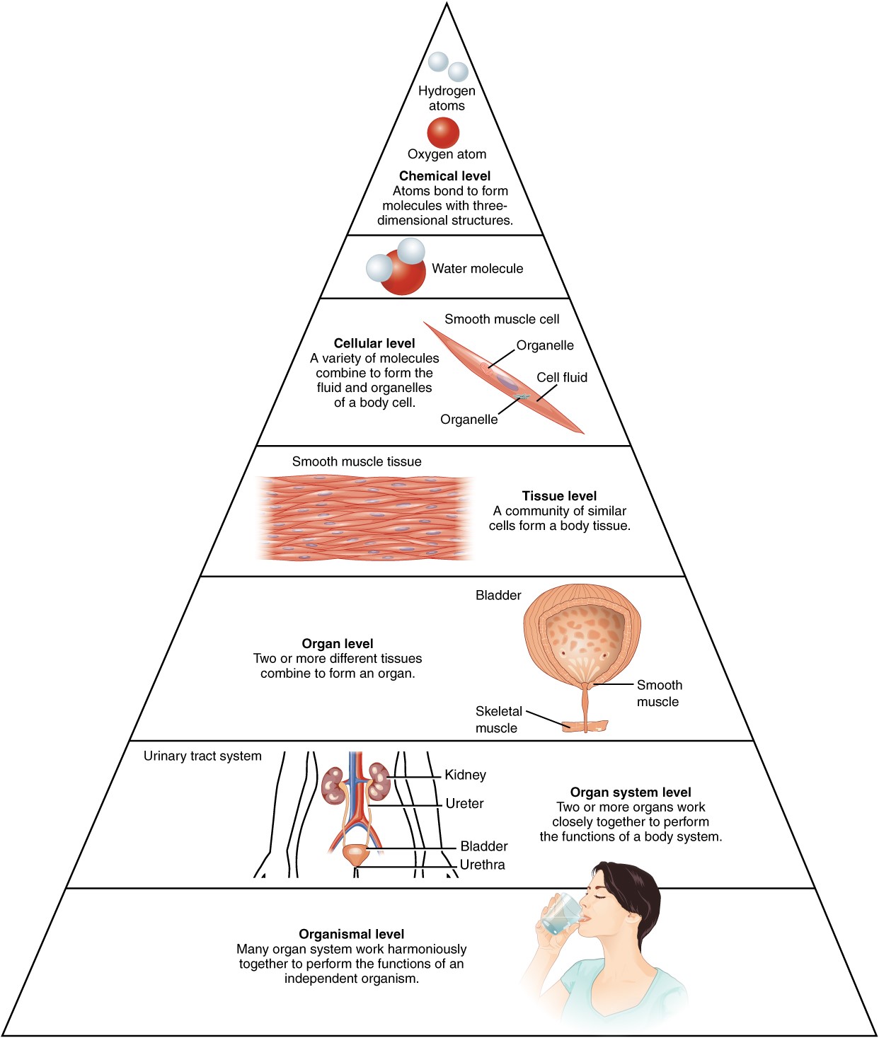
The chemical level of organization includes the simplest building blocks of matter: subatomic particles, atoms and molecules. Subatomic particles (protons, neutrons, and electrons) combine to form atoms. Familiar examples of atoms include hydrogen, oxygen, carbon, nitrogen, calcium, and iron. Two or more atoms combine to form a molecule, which includes things like water molecules, proteins, and sugars found in living things. Molecules are the chemical building blocks of all body structures.
A cell is the smallest independently functioning unit of a living organism which can include independently living single cell organisms like bacteria. All living structures within the human body contain cells, and almost all functions of human physiology are performed in cells or are initiated by cells. A human cell typically consists of flexible membranes that enclose cytoplasm, a water-based cellular fluid together with a variety of tiny functioning units called organelles. A tissue is a group of multiple similar cells (these cells can either be of the same cell type or can consist of a few related cell types) that work together to perform a specific function. An organ is an anatomically distinct structure of the body composed of two or more tissue types that performs one or more specific functions. An organ system is a group of organs that work together to perform major functions to meet physiological needs of the body. Throughout this course we will cover a subset of the organ systems found in the human body: the integumentary, skeletal, muscular, and nervous systems.
Language of Anatomy
Anatomists and health care providers use terminology to precisely talk about the anatomy of the human body that can seem overwhelming at first. The purpose of this language is not to confuse, but rather to increase precision, efficiency, and to reduce medical errors. For example, if you tell a friend that you have a scar “above the wrist” is it located on the forearm two or three inches away from the hand? Or is it at the base of the hand? Is it on the palm-side or back-side? By using precise anatomical terminology, including anatomical position, regional terms, directional terms, body planes, and body cavities, we can eliminate ambiguity and increase precision.
Anatomical terms are made up of roots, prefixes, and suffixes. The root of a term often refers to an organ, tissue, or condition, whereas the prefix or suffix often describes the root. For example, in the disorder hypertension, the prefix “hyper-” means “high” or “over,” and the root word “tension” refers to pressure, so the word “hypertension” refers to abnormally high blood pressure.
Anatomical Position
Anatomists have standardized the position of the body when it is referenced using descriptive terms to increase precision in language. Just as maps are normally oriented with north at the top, the standard body “map,” called anatomical position, is that of the body standing upright, with the feet at shoulder width and parallel, toes forward. The upper limbs are held out to each side, and the palms of the hands face forward (see Figures 1.3 or 1.4 for an example). Using this standard position helps reduce confusion and increase precision while describing parts of the human body. It does not matter how the body being described is oriented (ex: a doctor describing their patient who is sitting on an exam table), the terms are used as if that person is in anatomical position. For example, a scar in the “anterior (front) carpal (wrist) region” would always be present on the palm side of the wrist. The term “anterior” would always be used even if the hand were palm down on a table.
A body that is lying down is described as either prone or supine. Prone describes a face-down orientation, and supine describes a face up orientation. These terms are sometimes used in describing the position of the body during specific physical examinations or surgical procedures and you may hear the terms used to describe the position of the cadavers used in this course.
Regional Terms
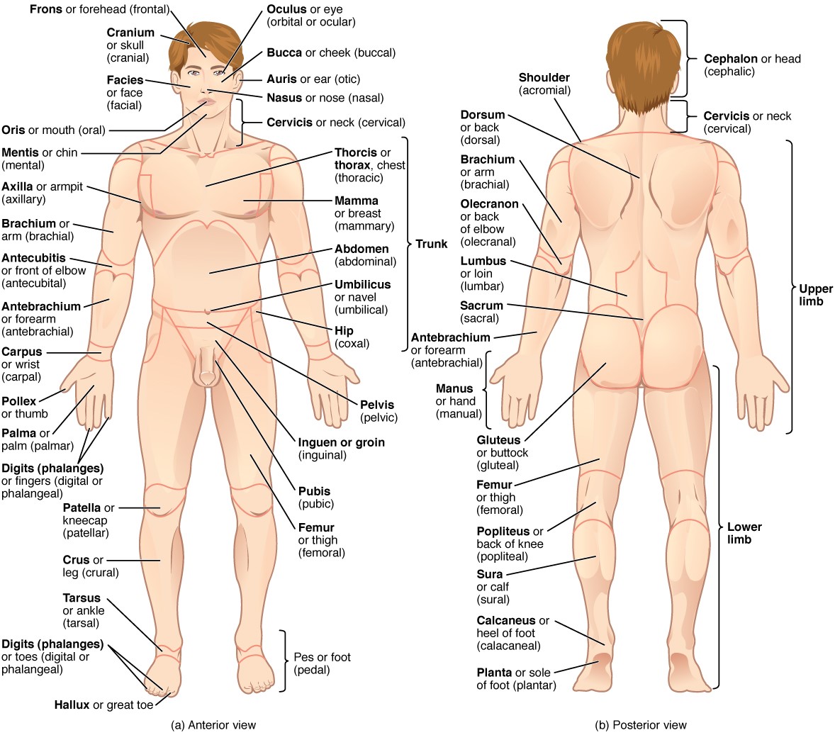
The human body’s numerous regions have specific terms to help increase precision in language (see Figure 1.3). Notice that the term “brachium” or “arm” is reserved for the “upper arm” and “antebrachium” or “forearm” is used rather than “lower arm.” Similarly, “femur” or “thigh” is correct, and “leg” or “crus” is reserved for the portion of the lower limb between the knee and the ankle. You will see these terms throughout the semester as they often form the basis for many of the structures you will learn later.
Directional Terms
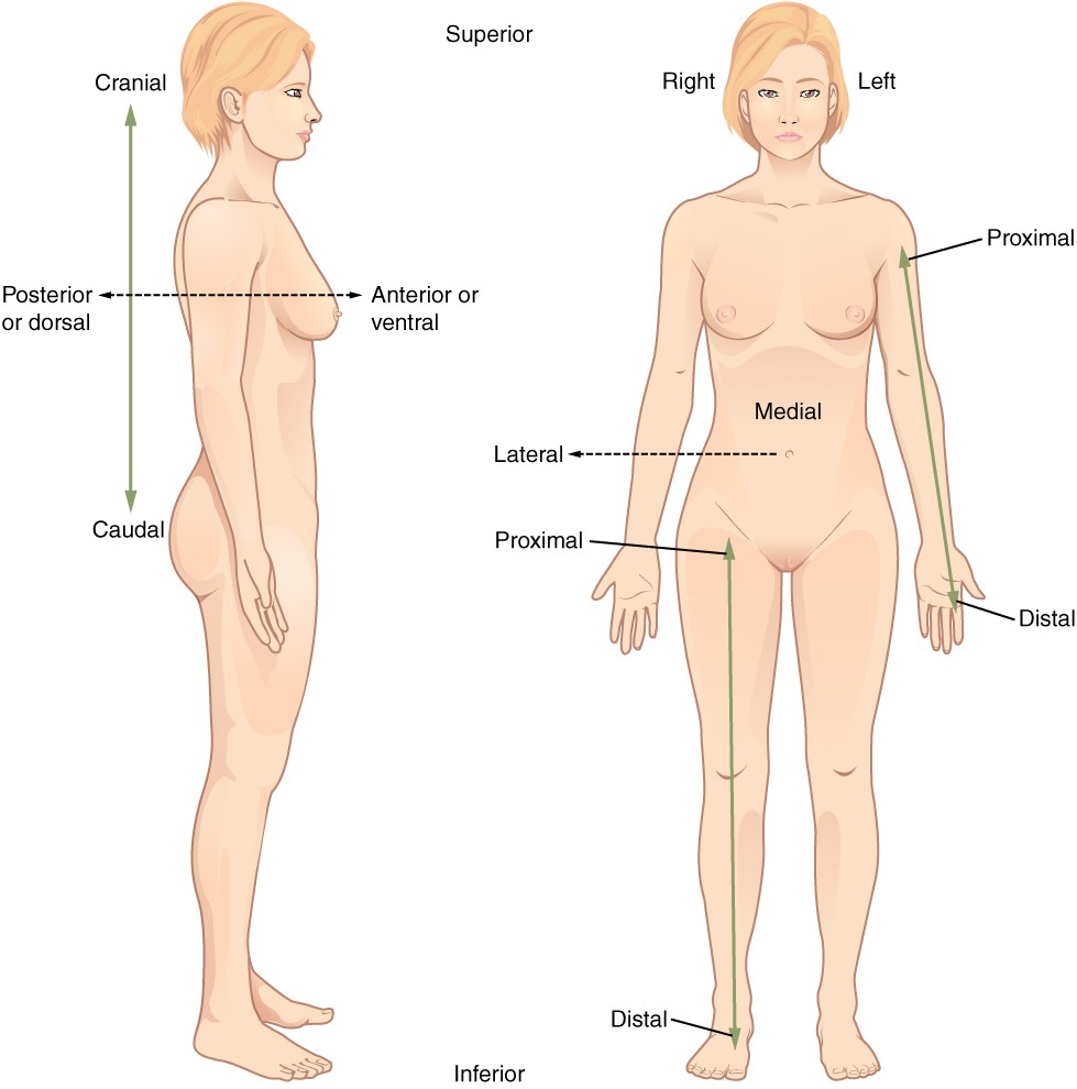
A set of specific directional anatomical terms appear throughout this and most other anatomy textbooks (Figure 1.4). These terms are essential for describing the relative locations of different body structures. For instance, an anatomist might describe one band of tissue as “inferior to” another or a physician might describe a tumor as “superficial to” a deeper body structure. Learning these terms now is critical to avoid confusion when you are studying or describing the locations of particular body parts in this course and in any future study of the human body.
- Anterior (or ventral) – Describes the front or direction toward the front of the body. For example, the toes are found on the anterior portion of the foot.
- Posterior (or dorsal) – Describes the back or direction toward the back of the body. For example, the spinal column is posterior to the sternum.
- Superior (or cranial) – Describes a position above or higher than another part of the body. For example, the eyes are superior to the mouth. Superior and cranial can often be used interchangeably though cranial is used to specifically refer to a structure near or toward the head. In quadrupeds the terms sometimes cannot be used interchangeably.
- Inferior (or caudal) – Describes a position below or lower than another part of the body. For example, the pelvis is inferior to the abdomen. Inferior and caudal can often be used interchangeably though caudal is used to specifically refer to a structure near or toward the tail (in humans, the coccyx, or lowest part of the spinal column). In quadrupeds the terms sometimes cannot be used interchangeably.
- Lateral – Describes the side or direction toward the side of the body. For example, the thumb is lateral to the other digits.
- Medial – Describes the middle or direction toward the middle of the body. For example, the big toe is the most medial toe.
- Proximal – Describes a position in a limb that is nearer to the point of attachment or the trunk of the body. For example, the upper arm is proximal to the wrist.
- Distal – Describes a position in a limb that is farther from the point of attachment or the trunk of the body. For example, the foot is distal to the thigh.
- Superficial – Describes a position closer to the surface of the body. For example, the skin is superficial to the bones.
- Deep – Describes a position farther from the surface of the body. For example, the brain is deep to the skull.
- Contralateral – Describes structures found on opposite sides of the body (right vs. left side). For example, the right foot is contralateral to the left arm.
- Ipsilateral – Describes structures found on the same side of the body. For example, the right hand and right shoulder are ipsilateral.
Body Sections & Planes
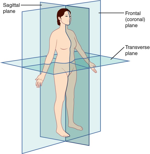
A section is a two-dimensional surface of a three-dimensional structure that has been cut. Modern medical imaging devices enable clinicians to obtain “virtual sections” of living bodies which we call these scans. Body sections and scans can be correctly interpreted, however, only if the viewer understands the plane along which the section was made. A plane is an imaginary two-dimensional surface that passes through the body. There are three planes commonly referred to in anatomy and medicine (Figure 1.5).
- Sagittal plane – Divides the body or an organ vertically into right and left sides. If this vertical plane runs directly down the middle of the body, it is called the midsagittal or median plane. If it divides the body into unequal right and left sides, it is called a parasagittal plane.
- Frontal plane – Divides the body or an organ into an anterior (front) portion and a posterior (rear) portion. The frontal plane is sometimes referred to as a coronal plane.
- Transverse plane – Divides the body or organ horizontally into upper and lower portions. Transverse planes produce images referred to as cross sections.
Body Cavities
The body maintains its internal organization by means of membranes, sheaths, and other structures that separate compartments. The dorsal (posterior) cavity and the ventral (anterior) cavity are the largest body compartments (Figure 1.6). These cavities contain delicate internal organs, and the ventral cavity allows for significant changes in the size and shape of the organs as they perform their functions. The lungs, heart, stomach, and intestines, for example, can change their shape considerably during expansion or contraction without distorting other tissues or disrupting the activity of nearby organs since they are found in cavities.
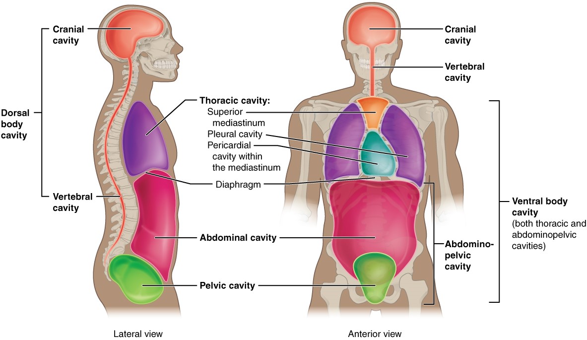
The dorsal and ventral cavities are each subdivided into smaller cavities. In the dorsal cavity, the cranial cavity houses the brain, and the vertebral (spinal) cavity encloses the spinal cord. Just as the brain and spinal cord make up a continuous, uninterrupted structure, the cranial and spinal cavities that house them are also continuous. The brain and spinal cord are protected by the bones of the skull and vertebral column and by cerebrospinal fluid, a colorless fluid produced by the brain, which cushions the brain and spinal cord within the dorsal cavity.
The ventral cavity has two main subdivisions: the thoracic cavity and the abdominopelvic cavity. The thoracic cavity is the more superior subdivision of the anterior cavity, and it is enclosed by the rib cage. The thoracic cavity contains the lungs (each found in a pleural cavity) and the heart (found in a pericardial cavity). The diaphragm forms the floor of the thoracic cavity and separates it from the more inferior abdominopelvic cavity. The abdominopelvic cavity is the largest cavity in the body. Although no membrane physically divides the abdominopelvic cavity, it can be useful to distinguish between the abdominal cavity, the division that primarily houses the digestive organs, and the pelvic cavity, the division that primarily houses the organs of reproduction.
Abdominal Regions and Quadrants
Health care providers typically divide up the abdominal cavity into either nine regions or four quadrants in order to promote clear communication about the location of a patient’s symptoms such as abdominal pain or a suspicious mass (Figure 1.7).
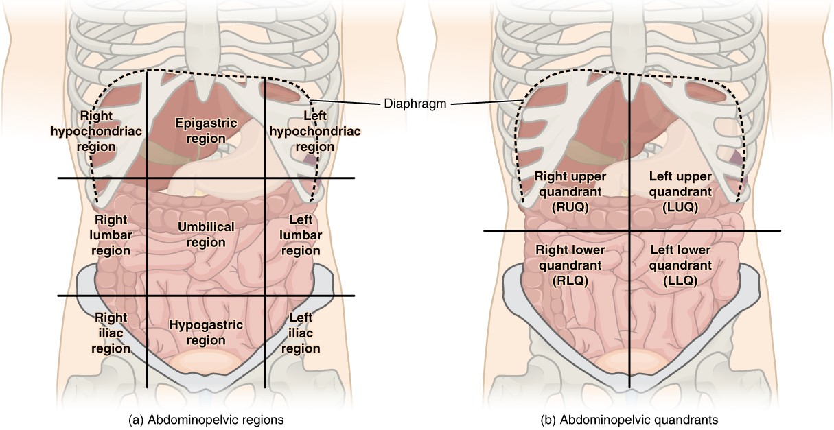
The more detailed regional approach subdivides the cavity with one horizontal line immediately inferior to the ribs and one immediately superior to the pelvis, and two vertical lines drawn as if dropped from the midpoint of each clavicle (collarbone). There are nine resulting regions. The simpler quadrants approach, which is more commonly used in medicine, subdivides the cavity with one horizontal and one vertical line that intersect at the patient’s umbilicus (navel).
Pre-Laboratory Questions
After you review the background information in this chapter answer the following questions prior to attempting the exercises.
- List the levels of organization in living things starting from the simplest to the most complex.
- What is the difference between anatomy and physiology?
- List the main regional terms used to indicate body regions in anatomy. Which body part does each represent?
- What are the four terms used to indicate the body planes or sections in anatomy? Describe each.
- List the main body cavities. Indicate the types of organs found in each.
- What are the terms used for the four abdominal quadrants and nine abdominal regions?
Exercises
- Exercise 1. Describe the hierarchy of organization of the human body
- Exercise 2. Demonstrate and describe anatomical position
- Exercise 3. Use directional terms to precisely describe the location of structures on the human body
- Exercise 4. Demonstrate and describe anatomical planes of section
- Exercise 5. Identify the major body cavities and provide examples of major organs found in each
Exercise 1 Describe the hierarchy of organization of the human body
Required Materials
• None
Procedure
This activity will be completed individually or in small groups. Refer to the background information to answer the questions below.
Complete the table below by sorting the given organizational levels of the human body from smallest to largest and then providing a one-sentence definition of each level.
Tissue; organelle; atom; organ; organ system; cell; organism; molecule
|
Smallest |
Definition |
|
|
|
|
|
|
|
|
|
|
|
|
|
|
|
|
|
|
|
|
|
|
|
|
|
Largest |
|
Exercise 2 Demonstrate and describe anatomical position
Required Materials
- A lab partner
- Open space
Procedure
Using the definition of anatomical position provided in the background information, take turns with a classmate to give simple, one-movement verbal instructions to transition from the given starting positions so that they end up in anatomical position.
Starting Positions:
- Lying face-up on the ground with their head, back, hands, and feet on the floor with both knees bent
- In a seated position on the floor with their legs straight and arms folded across their chest
- Sitting in a chair with their back to you and hands sitting in their lap
- Standing and facing you with their legs crossed and hands in their pocket
Check Your Understanding
Write your detailed step-by-step instructions in the provided table.
|
Scenario |
Given Instructions |
|
1 |
|
|
2 |
|
|
3 |
|
|
4 |
|
Exercise 3 Use directional terms to precisely describe the location of structures on the human body
Required Materials
- Post-its
- Skeleton or torso model
Procedure
This activity will be completed individually or in small groups. Use all of the directional terms provided in the table below in an accurate context by illustrating the terms on a skeleton or torso model.
Complete the table below for each directional term.
|
Directional Term |
Definition |
Example(s) |
|
Superior (cranial) |
|
|
|
Inferior (caudal) |
|
|
|
Medial |
|
|
|
Lateral |
|
|
|
Superficial |
|
|
|
Deep |
|
|
|
Anterior (ventral) |
|
|
|
Posterior (dorsal) |
|
|
|
Proximal |
|
|
|
Distal |
|
|
|
Ipsilateral |
|
|
|
Contralateral |
|
|
Exercise 4 Demonstrate and describe anatomical planes of section
Required Materials
- One pickle
- Plate
- Knife
- 4 Toothpicks
- Piece of paper
Procedure
This activity will be completed together as a class. Please do not eat the pickles.
- Retrieve a pickle on a plate, four toothpicks, and a knife from your instructor.
- Place the toothpicks in your pickle to serve as representations of the arms and legs.
- Your instructor will direct you to cut your pickle along one of five planes: midsagittal (median), parasagittal, frontal (coronal), transverse (horizontal), and oblique.
- Draw a representation of the now-visible section where you made the cut on your piece of paper.
- Compare your drawing and pickle-sections with other groups that made that same section.
- View the drawings of other groups that made different sections.
- Take a picture of your drawing and a representative example of each of the other four sections from other groups in the class.
For each of the following questions there could be one or more than one correct answer.
Choose the body plane(s) that would allow you to see both lungs at the same time:
Midsagittal
Parasagittal
Frontal
Transverse
Oblique
Choose all possible body plane(s) that would allow you to see the brain and the spinal cord:
Midsagittal
Parasagittal
Frontal
Transverse
Oblique
Choose the body plane(s) that would allow you to see the brain but not the spinal cord:
Midsagittal
Parasagittal
Frontal
Transverse
Oblique
Choose the body plane(s) that would allow you to see the right eye but not the left eye:
Midsagittal
Parasagittal
Frontal
Transverse
Oblique
Exercise 5 Identify the major body cavities and provide examples of major organs found in each
Required Materials
- Post-its
- Large piece of paper
- Tape
- Torso model and/or a classmate
Procedure
This activity will be completed as a group.
On the large piece of paper, draw two perpendicular lines to create four quadrants (right-upper, right-lower, left-upper, and left-lower), similar to Figure 1.7.
Tape the piece of paper onto the abdomen of the torso model or a classmate
Your instructor will call out the name of a major organ and you will write the organ name on a post-it and then place the post-it in the correct quadrant.
List all of the cavities found within the dorsal body cavity.
List all of the cavities found within the ventral body cavity.
Complete the table to provide one example of an organ found in each of the following body cavities.
|
Body cavity |
Organ Example(s) |
|
Cranial |
|
|
Abdominal |
|
|
Pelvic |
|
|
Pleural |
|
|
Vertebral |
|
|
Pericardial |
|
1.Name the six levels of organization of the human body.
a.
b.
c.
d.
e.
2.Describe the anatomic position using your own words?
3. If you use a midsagittal section, which organs can you divide into two equal sections? Give at least one example and demonstrate this sectioning by using a sketch of the organ.
4. Fill in the gaps below using an appropriate directional term.
- The head is located __________ to the toes.
- In the anatomic position, the thumb is located _______ to the pinky of the same hand.
- The mouth is located ________ to the eyes.
- Muscles lie ______ to the skin covering in their area.
- The tip of your nose is ______ to the top of your ears.
5. Examine the figures above and do some additional research to list all the organs that are within each of the four abdominal quadrants. Sketch and show the quadrants and the organs contained within each.
6. Make a list of the body cavities of the human body. Then, list all the organs that are found within each cavity.
7. In anatomical terminology we use specific words to define each body region. List the regional term that is used for each of the following every day language terms we use to describe these body regions:
- head
- neck
- face
- eye
- ear
- nose
- mouth
- arm
- armpit
- wrist
- finger
- chest
- breast
- thigh
- kneecap
- back of knee
- ankle
- back
- navel
- skull
