
17 Chapter 17 The Endocrine System
By Rajeev Chandra
Motivation.

You may never have thought of it this way, but when you send a text message to two friends to meet you at the dining hall at six, you’re sending digital signals that (you hope) will affect their behavior—even though they are some distance away. Similarly, certain cells send chemical signals to other cells in the body that influence their behavior. This long-distance intercellular communication, coordination, and control is critical for homeostasis, and it is the fundamental function of the endocrine system.
The laboratory exercises in this module will identify several of the “classical” endocrine glands, describe the location and structure of these glands (both gross and histological), and outline the functions of the hormones that are secreted from these glands. Regulation of the glands by factors in the internal environment will also be explored in a simulated experiment.
Learning Objectives
Upon completion of the work in this chapter students should be able to:
- Identify the classical endocrine glands on a model or diagram
- List the hormones produced by each endocrine gland identified, and discuss the actions of each hormone identified
- Differentiate among the histology of the endocrine glands when viewed on a microscope slide
- Identify the zona glomerulosa, zona fasciculata, zona reticularis, and adrenal medulla, and list the hormones that are released from each region
- Identify pancreatic islets
- Describe the nature of the feedback loops that regulate the activity of the hypothalamus, pituitary gland, the target glands, and target tissues
Background.
Overview
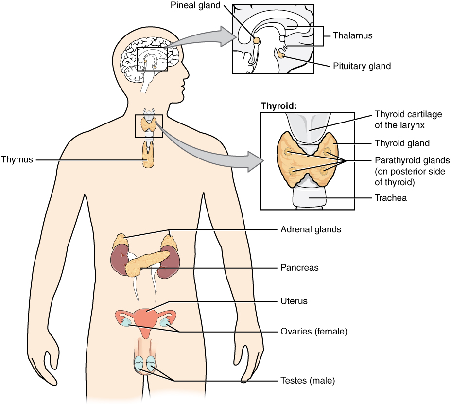
Hormones are chemical messengers produced by cells and released into the bloodstream, where they travel throughout the body. This allows these messengers to act on cells and organs that may be distant from the cells that synthesized them. Although many cells release chemicals that communicate with other cells, we typically describe some organs or glands as being part of the “endocrine system.” Glands that are typically included in discussions of the anatomy and physiology of the endocrine system are shown at right.
Each endocrine gland secretes particular hormones, which in turn has particular effects on the target cells and organs. We’ll be looking at these glands and hormones individually as part of this exercise.
SPECIFIC ENDOCRINE GLANDS
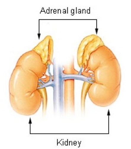
Adrenal glands
The adrenal glands (as their name implies) are located near the kidneys (ad-RENAL). They are paired, pyramidal glands that sit on the superior aspect of each kidney.
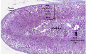
The adrenal glands are composed of two distinct regions: the outer adrenal cortex, and the inner adrenal medulla. These regions can be seen when the glands are sectioned, but the real differences are clear when the tissue is viewed under a microscope.
Adrenal Cortex
The adrenal cortex can be further divided into 3 sub-regions or layers:
The zona glomerulosa, the outermost or most superficial layer, which synthesizes and secretes aldosterone in response to stimuli like decreased blood pressure. Aldosterone
(a steroid) regulates Na+ and K+ balance, and plays an important role in fluid homeostasis.
The zone fasciculata, the middle layer, synthesizes and secretes cortisol and other glucocorticoids in response to ACTH from the anterior pituitary.
The zona reticularis, the deepest layer of the cortex, secretes androgens, or sex hormones (that are also steroids). These hormones act throughout the body and have similar effects as sex steroids produced by the ovaries and testes.
Adrenal Medulla
The adrenal medulla is the innermost (deepest) region of the adrenal gland, and consists mostly of modified sympathetic nerves. As a result, the adrenal medulla synthesizes and secretes catecholamines epinephrine and norepinephrine into the bloodstream.
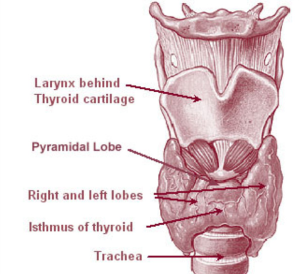
Thyroid gland
The thyroid gland is a butterfly-shaped gland located on the anterior aspect of the larynx (voicebox). The right and left lobes on either side (analogous to the butterfly’s wings) are connected by a thin band of glandular tissue called the isthmus (Figure 17.5).
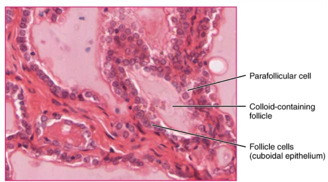
Histologically, the thyroid gland is composed of follicles, round structures where thyroid hormones are synthesized and released. Several follicles can be seen in the photomicrograph below. The substance in the follicles is called colloid, which is an iodine-rich precursor to thyroid hormones. The follicular cells use these precursor molecules to synthesize and release the finished thyroid hormones (triiodothyronine, or T3, and thyroxine, or T4). Thyroid hormones act on many other cells in the body and have many effects, including increasing heart rate, increasing protein synthesis, and increasing overall metabolic rate. Parafollicular cells are found in-between the follicles. These cells synthesize another hormone called calcitonin, which plays a role in calcium balance in the body.
Parathyroid glands
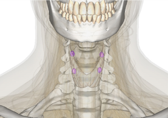
The parathyroid glands are 4 – 6 small, round glands found on the posterior aspect of the thyroid gland (Figure 17.7). These glands secrete parathyroid hormone (PTH) in response to low levels of calcium in the plasma. PTH acts to activate osteoclasts to dissolve existing bone tissue, liberating the calcium that is stored there. PTH also acts to increase calcium absorption from the intestines and increases the reabsorption of calcium in the kidney tubules. The net result of PTH on the body is to increase calcium ion levels in the plasma.
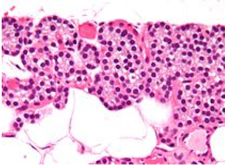
A photomicrograph of the parathyroid gland (Figure 17.8) shows the cells that are responsible for the synthesis and release of PTH called chief cells, or parathyroid cells. Other cells that are present in the parathyroid glands are called oxyphil cells, but their function is not clear.
Pancreas
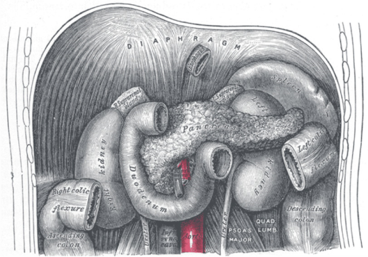
The pancreas is a large, comma-shaped organ (Figure 17.9) that has both exocrine functions (synthesizing digestive juices that are secreted into the small intestine) and endocrine functions. It is located in the abdominal cavity, posterior to the stomach and tucked into the curve of the duodenum.
Several hormones are synthesized and secreted from the pancreas, including insulin, glucagon, and somatostatin. (Somatostatin is known by another name – growth hormone inhibiting hormone, the same hormone that is released from the hypothalamus to regulate the activity of the pituitary gland.)
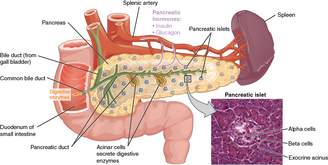

The two main hormones released from the pancreatic islet cells are insulin and glucagon. Insulin is released from beta cells in the pancreatic islets, and has the actions to increase the uptake of glucose from the blood into tissues. This results in decreased glucose in the blood plasma. Glucagon is released from alpha cells, and has the effect to release glucose from body cells into the plasma, resulting in increased blood glucose levels. Somatostatin is released from delta cells in the islets. The action of somatostatin is generally inhibitory. In the digestive system, somatostatin acts to inhibit the release of both insulin and glucagon. In the anterior pituitary (discussed above), somatostatin inhibits the release of growth hormone (GH) from the anterior pituitary.
Reproductive Glands: Ovaries and Testes
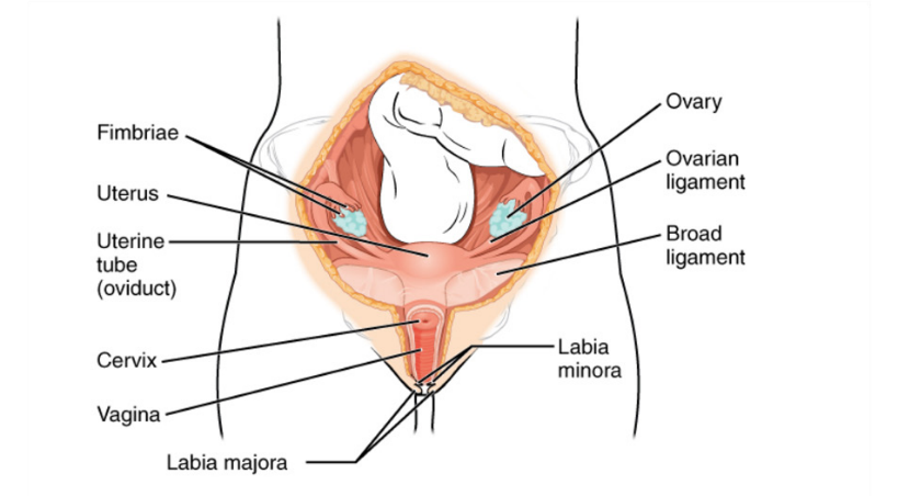
The ovaries (Figure 17.12) are the female gonads that produce both gametes (cells that play a role in reproduction) and hormones. They are small, paired glands located deep in the pelvic cavity, and the gametes they produce are called oocytes. The production of oocytes is dependent on the hormonal activity of the anterior pituitary. The oocytes are released from the ovary and travel through the uterine tube to the uterus.
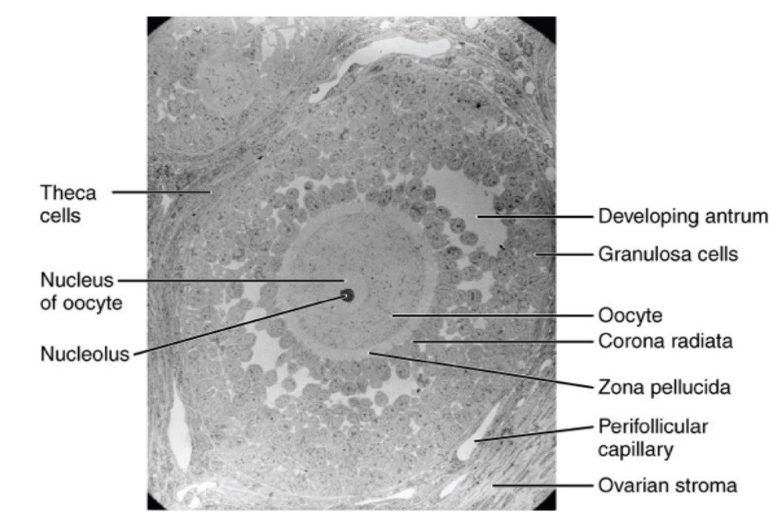
Ooctyes develop within follicles in the cortex of the ovary (Figure 17.13). The process where oocytes are released from the ovary is called ovulation.
The ovary also produces female sex hormones called estrogens. There are three estrogens produced by the ovary: estrone, estradiol and estriol. All the estrogens are classified as steroids due to their chemical structure. Estrogens regulate the ovarian and menstrual cycles, influencing the maturation of the oocyte in the ovary, the release of the oocyte during ovulation, and the changes seen in the uterus as it prepares to receive a potential zygote. Estrogens are also important in the development of secondary sex characteristics like breast development and fat redistribution. Another hormone produced by the ovary is progesterone, a hormone that plays important roles in preparing the body for pregnancy in the event that an oocyte is fertilized by a sperm cell.
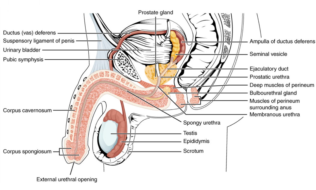
The testes are male gonads (Figure 17.14) , and like the ovaries in females they produce both gametes (sperm) and hormones. The testes are also small paired structures, however they are located outside the pelvic cavity in males. Sperm that are produced in the testes must travel back into the pelvic cavity through a long duct system before being released from the body via the penis as part of the semen. The production of sperm in the male is regulated similarly to the oocyte production in females, via hormones that are released from the anterior pituitary.
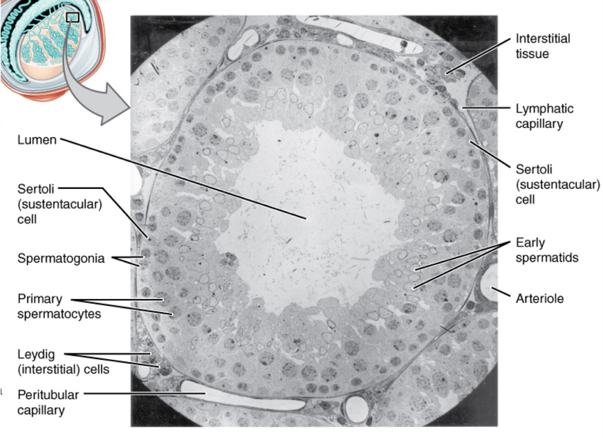
Sperm is produced within the seminiferous tubule (Figure 17.15). Maturing sperm move through an extensive duct system both in the testis and to / through the pelvic cavity.
The main male sex hormone produced by the testes is testosterone. Like the estrogens from the ovary, testosterone is a steroid hormone that plays important roles in the development of secondary sex characteristics in males (increased bone and muscle mass, deepening voice, facial hair). Testosterone also influences the development of the sperm. A second hormone produced by the testes is inhibin, which plays a role in regulating the development and maturation of sperm.
Pituitary Gland and Hypothalamus
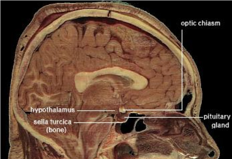
The pituitary gland extends below the inferior aspect of the brain, inferior to the hypothalamus (Figure 17.16). We describe the pituitary gland as the “master gland” because it secretes hormones that regulate the activity of other endocrine glands, in addition to hormones that act on other cells and tissues in the body.
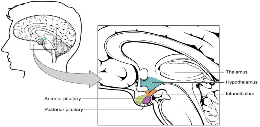
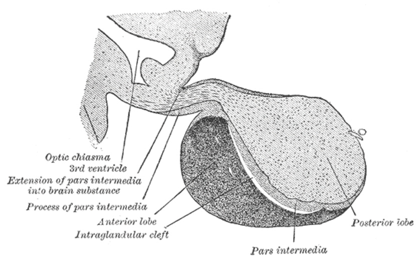
Pituitary Complex The hypothalamus region lies inferior and anterior to the thalamus (Figure 17.17). It connects to the pituitary gland by the stalk-like infundibulum. The pituitary gland consists of an anterior and posterior lobe, with each lobe secreting different hormones in response to signals from the hypothalamus. The pituitary gland is composed of 2 distinct regions, or lobes: the ANTERIOR lobe (also called the adenohypophysis; sometimes called the pars distalis) and the POSTERIOR lobe (also called the neurohypophysis; sometimes called the pars nervosa).The differences between the two lobes can be seen easily when the tissue is examined under the microscope.
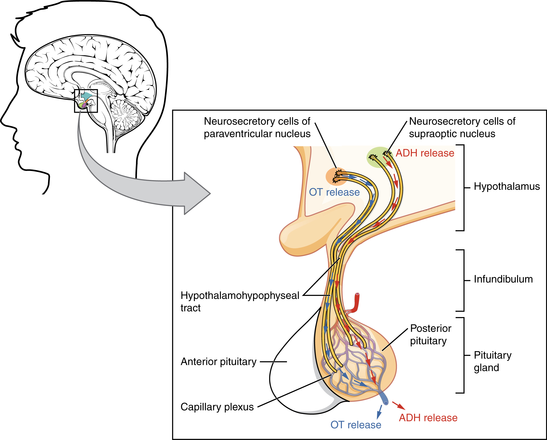
The posterior lobe is an extension of the brain tissue in the hypothalamus. This means that the posterior lobe is actually nervous tissue and not true glandular tissue. Hormones are synthesized in cell bodies in the hypothalamus, then transported to the axon terminals in the posterior lobe. These hormones are released from the axon terminals into the bloodstream. The anterior lobe is true glandular tissue: hormones are synthesized, stored and released from the cells here.
Hormones Secreted from the Pituitary Gland
Two hormones are secreted from the posterior pituitary:
- Antidiuretic hormone (ADH)
- Oxytocin
Neurosecretory cells in the hypothalamus release oxytocin (OT) or ADH into the posterior lobe of the pituitary gland. These hormones are stored or released into the blood via the capillary plexus (Figure 17.19).
Six hormones are released from the anterior pituitary:
- Growth hormone (GH)
- Prolactin
- Follicle-stimulating hormone (FSH)
- Luteinizing hormone (LH)
- Adrenocorticotropic hormone (ACTH)
- Thyroid-stimulating hormone (TSH)
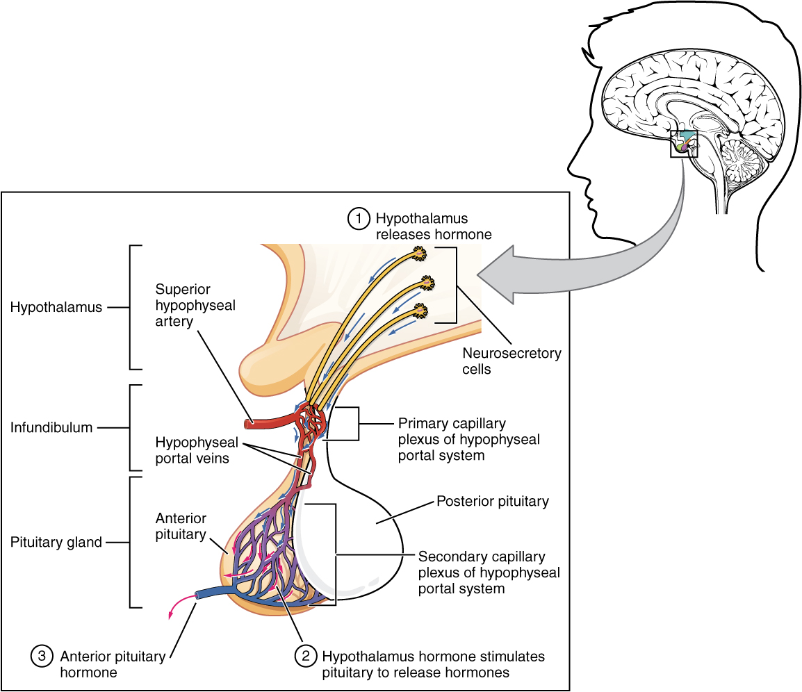
The anterior pituitary is composed of three distinct cell types that can be seen more easily when they are stained with acidic or basic dyes. ACIDOPHILS are cells that appear red when stained with acidic dyes. BASOPHILS are cells that appear blue when stained with basic dyes. CHROMOPHOBES are cells that remain uncolored in the presence of either type of dye. The hormones that are released from the anterior pituitary are associated with either acidophils or basophils. The table below (Post-Laboratory Questions, #4) shows you which hormones are released from each type of cell.
REGULATION OF THE PITUITARY AND TARGET ORGANS / GLANDS
Feedback Loops
The release of hormones in the endocrine system are regulated through feedback mechanisms. Negative feedback is used to inhibit further hormone secretion: when a sufficient amount of hormone is released, it “feeds back” to decrease or prevent further release. In other words, the gland has released sufficient hormone to produce the desired effect, so further hormone release is inhibited. Because the hypothalamus and pituitary glands are regulating the activity of endocrine glands, they are also subject to feedback mechanisms and their activity will be altered in response to levels of other hormones.
The regulation of the anterior pituitary involves the release of hormones from the hypothalamus; regulation of the endocrine glands involves the release of hormones from the anterior pituitary. Further, hormones released from the endocrine glands interact with both the hypothalamus and the pituitary to regulate their activity, in a classic arrangement known as a feedback loop.
The pathways of three hormones are examined in this activity: thyroid hormone, cortisol and testosterone. The hormonal pathways are similar in all three cases. In each case: the hypothalamus secretes a releasing hormone to regulate the activity of the anterior pituitary gland the anterior pituitary then secretes hormones that regulate the activity of a target gland (the thyroid gland, the adrenal gland or the testis) the hormones released from the target glands “feed back” to the anterior pituitary and the hypothalamus to inhibit the further release of those hormones. The hypothalamus is like a command center: if it is not stimulated, it will not secrete releasing hormones to stimulate the anterior pituitary, which in turn will not stimulate the target glands.
Pre-Laboratory Questions
After reviewing the Background information please answer the following questions.
- Endocrine glands ________.
- secrete hormones that travel through a duct to the target organs
- release neurotransmitters into the synaptic cleft
- secrete chemical messengers that travel in the bloodstream
- include sebaceous glands and sweat glands
- Chemical signaling that affects neighboring cells is called ________.
- autocrine
- paracrine
- endocrine
- neuron
- A student is in a car accident, and although not hurt, immediately experiences pupil dilation, increased heart rate, and rapid breathing. What type of endocrine system stimulus did the student receive?
- humoral
- hormonal
- neural
- positive feedback
- Which of the following is an anterior pituitary hormone?
- ADH
- oxytocin
- TSH
- Cortisol
- How many hormones are produced by the posterior pituitary?
- 0
- 1
- 2
- 6
- The adrenal glands are attached superiorly to which organ?
- thyroid
- liver
- kidneys
- hypothalamus
- The gonads produce what class of hormones?
- amine hormones
- peptide hormones
- steroid hormones
- catecholamines
- If an autoimmune disorder targets the alpha cells, production of which hormone would be directly affected?
- somatostatin
- pancreatic polypeptide
- insulin
- glucagon
Exercises
- Exercise 1 Identify major endocrine glands and their locations
- Exercise 2 Name the main hormones secreted by each major endocrine gland (optional)
- Exercise 3 Describe the function of the main hormones (optional)
- Exercise 4 Identify microscopic structures of endocrine glands
Exercise 1 Identify major endocrine glands and their locations
Required Materials
- Torso Model
- Organs of the Endocrine System board model
- The Endocrine System poster
- Post-it notes
- Labeling tape
Procedure
- This activity requires students identifying the locations of various Endocrine Glands using Torso Models. Students will also be able to explore respective structures of the Endocrine Glands using board model depicting various endocrine glands.
- Use the board model to identify and become familiar with the anatomy of the following endocrine glands: pituitary gland, thyroid gland, parathyroid glands, pancreas, adrenal gland, testis, ovary, pancreas. Place a label on each (using post-it notes) and take a picture. Insert the labeled picture in the space below. Alternatively, you can sketch and label.

- Now that you know what these glands look like, find them on the torso model. Use the post-it notes to label them. Take a picture of the labeled glands on the torso model and insert in the space below. Alternatively, you can sketch and label.
Exercise 2 Name the main hormones secreted by each major endocrine gland (optional)
Required Materials
- Endocrine system poster
Procedure
Students will be utilizing this lab manual and OpenStax Human Anatomy and Physiology Text resources as well as the Endocrine System Posterto learn and identify hormones secreted by various endocrine glands. For each gland listed in the table below, find and list the hormones it produces.
| Gland | Hormones produced |
| Pituitary (posterior) | |
| Pituitary (anterior) | |
| Thyroid | |
| Parathyroid | |
| Pancreas | |
| Adrenal | |
| Ovary | |
| Testis |
Exercise 3 Describe the function of the main hormones (optional)
Required Materials
- None
Procedure
Students will be utilizing this lab manual and OpenStax Human Anatomy and Physiology Text resources to learn and identify functions of some important hormones in the table below.
| Hormones | Hormone name and function |
| Pituitary (posterior) hormones | 1.
2. |
| Pituitary (anterior) hormones | 1.
2. 3. 4. 5. 6. |
| Thyroid | 1.
2. |
| Parathyroid | 1. |
| Pancreas | 1.
2. 3. |
| Adrenal | 1.
2. 3. |
| Ovary | 1.
2. |
| Testis | 1. |
Exercise 4 Identify microscopic structures of endocrine glands
Required Materials
- Compound microscope
- Microscope lens paper
- Microscope lens solution
- Microscope immersion oil
- Slide of Adrenal Section (human) (Figure 17.4)
- Mammal Thyroid and Parathyroid Glands slide (Figure 17.6 thyroid, 17.8 parathyroid)
- Human Pancreas slide (Figure 17.11)
- Slide of Mammal Ovarian Follicles (Figure 17.13)
- Human Testis slide (Figure 17.15)
- Hypophysis (pituitary gland) slide (Figure 17.18)
Procedure
For each of the 6 endocrine gland slides listed above, follow these instructions:
- Ask your instructor for a prepared slide. Refer to the Figures indicated above for each slide to see what you expect to observe.
- Place it on the stage of microscope and bring it into focus using low power (4 x 10 magnification).
- Study tissue on slide. Move slide vertically and horizontally.
- Change the magnification to high power (10 x 10).
- Draw the tissues at low and high magnification to show the cell and tissue structure as best as you can. Label.
Adrenal
Thyroid
Parathyroid
Pancreas
Ovarian follicle
Testis
Hypophysis (pituitary gland)
Post-laboratory Questions
- Hormones of the Pancreas. Fill in the below table with the hormone that is released from each cell type found in the pancreatic islets. In the last column, briefly describe the actions of each hormone.
| Cell type | Hormone released | Actions |
| Alpha cells
|
||
| Beta cells
|
||
| Delta cells
|
- Sex Hormones. Fill in the below table with the hormone that is released from each gonad. Note that there is more than one hormone released from each gonad. In the last column, briefly describe the actions of each hormone.
| Gonad | Hormone released | Actions – on gametes and on other cells in the body |
| Testes
|
||
| Ovary
|
||
- Summary of Hormone Action. For each hormone in the left-hand column, fill in the table with the target cells or tissues on which each hormone acts; the effects of the hormones on those target cells or organs; and the stimulus for the release of each hormone.
| Thyroid and Parathyroid Hormones | |||
| Hormone | Stimulus for Release | Target Cells / Tissues | Effects |
| Thyroid Hormones
(T3 and T4)
|
|||
| Calcitonin
|
|||
| Parathyroid Hormone
|
- Source, Actions, and Hypothalamic Regulation of the Anterior Pituitary. Fill in the table with the actions of each hormone, and the name of the hypothalamic hormone(s) that regulate their release.
| Pituitary Gland | Cells | Hormones Produced | Action of Hormones | Hypothalamic releasing or inhibiting hormone |
| Anterior
(adenohypophysis)
|
Acidophils
|
GH
Prolactin
|
||
| Basophils | FSH
LH ACTH TSH
|
|||
| Chromophobes | NA | NA | NA | |
| Posterior
(neurohypophysis) |
Axon terminals
|
ADH | NA | |
| Axon terminals
|
Oxytocin | NA | ||
