
6 Chapter 6 Bone Tissue and the Skeletal System
By Ganesan L. Kamatchi
Motivation.
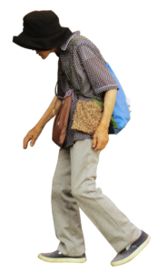
Healthy bones require good nutrition and lifestyle as well as a genetic component. Some bone diseases such as osteoporosis disproportionately affect some populations. Osteoporosis is a condition that reduces bone density by making the bones more porous or spongy. Women are more severely and frequently affected by this condition due to the dependence of bone producing and bone remodeling cells on a good balance of female hormones such as estrogen that decline over time. Alcoholism, smoking, anorexia, being of European heritage, surgical removal of ovaries, and some medications can increase risk of this painful condition called osteoporosis. Bone fractures that result from weakening of bones can be debilitating. A good diet, exercise, stopping alcohol and smoking, and some medications that help bone growth can be used for treatment of osteoporosis; but aging can’t be reversed!
Figure 6.1 Elderly woman with osteoporosis showing a curved back from compression fractures of her back bones. (Credit: Wikipedia. James Hailman, MD own work, CC-BY SA license)
Learning Objectives
Upon completion of the work in this chapter students should be able to:
- Demonstrate classification of bones based on shape and size
- Label the regions of long bone gross anatomy
- Sketch and describe the microscopic anatomy of compact and spongy bone
- Describe the development of long bones based on microscopic observation
Background.
Your skeleton is a structure of living tissue that grows, repairs, and renews itself. The bones within it are dynamic and complex organs that serve several important functions, including some necessary to maintain homeostasis. Bone, or osseous tissue, is a hard, dense connective tissue that forms most of the adult skeleton, the support structure of the body. In the areas of the skeleton where bones move (for example, the ribcage and joints), cartilage, a semi-rigid form of connective tissue, provides flexibility and smooth surfaces for movement.
The skeletal system is the body system composed of bones and cartilage and performs the following critical functions for the human body:
- Supports the body
- Facilitates movement
- Protects internal organs
- Produces blood cells
- Stores and releases minerals and fat
Classification of Bones
The 206 bones that compose the adult skeleton are divided into five categories based on their shapes (Table 6.1). Their shapes and functions are related such that each categorical shape of bone has a distinct function.
Table 6.1: Classification of Bones Based on their Shape
| Class of Bone | Features | Function | Examples |
| Long | Cylinder-like shape, longer than it is wide | Leverage | Femur, tibia, fibula, metatarsals, humerus, ulna, radius, metacarpals, phalanges |
| Short | Cube-like shape, approximately equal in length, width, and thickness | Provide stability, support, while allowing for some motion | Carpals, tarsals |
| Flat | Thin and curved | Points of attachment for muscles; protectors of internal organs | Sternum, ribs, scapulae, cranial bones |
| Irregular | Complex shape | Protect internal organs | Vertebrae, facial bones |
| Sesamoid | Small and round; embedded in tendons | Protect tendons from compressive forces | Patellae |
Gross Anatomy of Bone
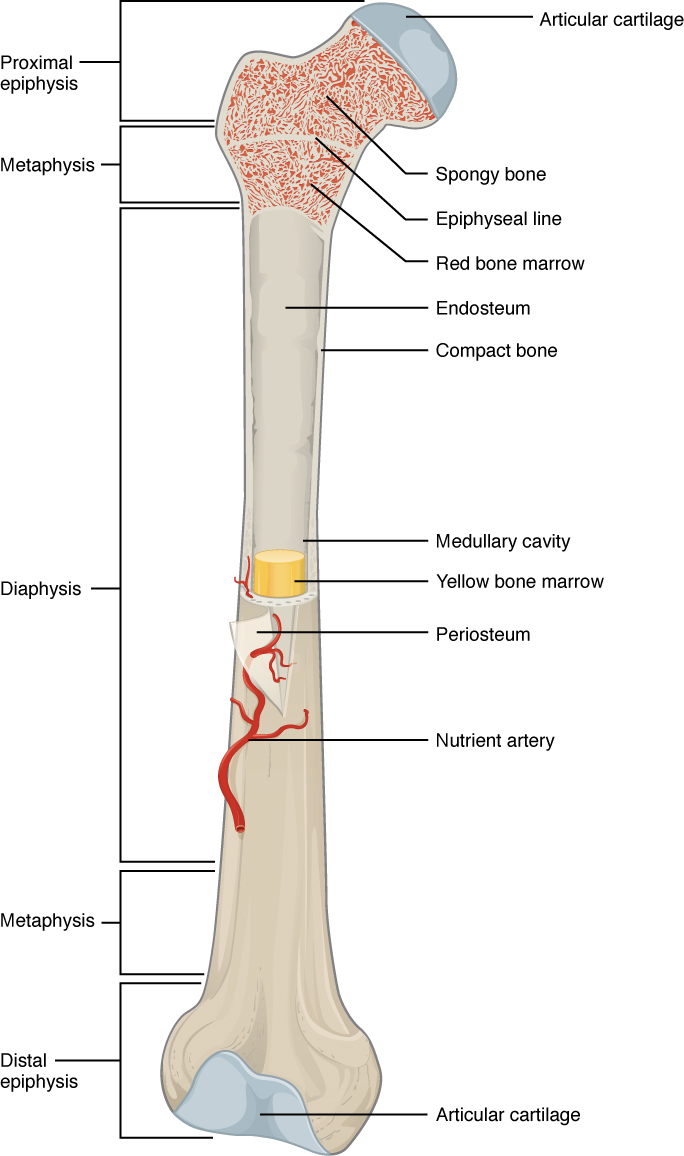
The structure of a long bone allows for the understanding of the gross anatomy of bone. A long bone has two parts, the long tubular shaft called diaphysis and the two wider ends, the epiphysis (Figure 6.2). The hollow region in the diaphysis is called the medullary cavity, which is filled with yellow marrow. The walls of the diaphysis are composed of dense and hard compact bone.
The epiphysis is filled with spongy bone and the space in the spongy bone is filled with red marrow. Each epiphysis meets the diaphysis at the metaphysis, the narrow area that contains the epiphyseal plate (growth plate).
The medullary cavity has a delicate membranous lining called the endosteum, where bone growth, repair, and remodeling occur. The outer surface of the bone is covered with a fibrous membrane called the periosteum which contains blood vessels, nerves, and lymphatic vessels. Tendons and ligaments also attach to bones at the periosteum. The periosteum covers the entire outer surface except where the epiphyses meet other bones to form joints.
Compact Bone
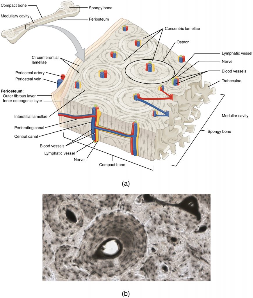
Compact bone is the denser, stronger of the two types of bone tissue. The microscopic structural unit of compact bone is called an osteon, or Haversian system. Each osteon is composed of concentric rings of calcified matrix called lamellae. Running down the center of each osteon is the central canal, or Haversian canal, which contains blood vessels, nerves, and lymphatic vessels. These vessels and nerves branch off at right angles through a perforating canal, also known as Volkmann’s canals, to extend to the periosteum and endosteum.
The osteocytes are located inside spaces called lacunae, found at the borders of adjacent lamellae. Canaliculi connect with the canaliculi of other lacunae and eventually with the central canal (Fig. 6.4). This system allows nutrients to be transported to the osteocytes and wastes to be removed from them.
Spongy Bone
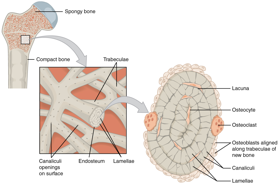
Spongy bone, also known as cancellous bone, contains osteocytes housed in lacunae, but they are not arranged in concentric circles. Instead, the lacunae and osteocytes are found in a lattice-like network of matrix spikes called trabeculae (Figure 6.4). The trabeculae may appear to be a random network, but each trabecula forms along lines of stress to provide strength to the bone. The spaces of the trabeculated network provide balance to the dense and heavy compact bone by making bones lighter so that muscles can move them more easily. In addition, the spaces in some spongy bones contain red marrow, protected by the trabeculae, where hematopoiesis occurs.
Bone Development
Long bones develop using endochondral ossification and flat bones develop using intramembranous ossification. Here we will focus on endochondral bone development
In endochondral ossification, bone develops by replacing hyaline cartilage. Cartilage serves as a template to be completely replaced by new bone. Endochondral ossification takes much longer than intramembranous ossification. Bones at the base of the skull and long bones form via endochondral ossification.
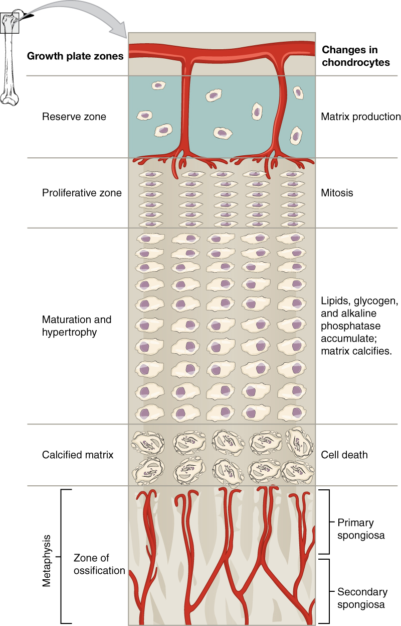
The epiphyseal plate is the area of growth in a long bone. It is a layer of hyaline cartilage where ossification occurs in immature bones. On the epiphyseal side of the epiphyseal plate, cartilage is formed. On the diaphyseal side, cartilage is ossified, and the diaphysis grows in length. The epiphyseal plate is composed of four zones of cells and activity (Figure 6.5). The reserve zone is the region closest to the epiphyseal end of the plate and contains small chondrocytes within the matrix.
The proliferative zone is the next layer toward the diaphysis and contains stacks of slightly larger chondrocytes. It makes new chondrocytes (via mitosis) to replace those that die at the diaphyseal end of the plate. Chondrocytes in the next layer, the zone of maturation and hypertrophy, are older and larger than those in the proliferative zone. The longitudinal growth of bone is a result of cellular division in the proliferative zone and the maturation of cells in the zone of maturation and hypertrophy.
Most of the chondrocytes in the zone of calcified matrix, the zone closest to the diaphysis, are dead because the matrix around them has calcified. Capillaries and osteoblasts from the diaphysis penetrate this zone, and the osteoblasts secrete bone tissue on the remaining calcified cartilage. Thus, the zone of calcified matrix connects the epiphyseal plate to the diaphysis. A bone grows in length when osseous tissue is added to the diaphysis.
Pre-Laboratory Questions
After you review the background information, please answer the following questions.
- What is the type of tissue, the bones made of?
- What are the types of bone cells and where do they come from?
- Describe the organization of osteon?
- Describe the parts of a bone?
- What are the functions of bone?
Exercises
- Exercise 1. Identification of Classes of Bones Based on Shape
- Exercise 2. Gross Anatomy of Bone
- Exercise 3. Compact Bone
- Exercise 4. Spongy Bone
- Exercise 5. Endochondral Bone Development
Exercise 1 Identification of Classes of Bones Based on Shape
Required Materials
- Disarticulated human skeleton
Procedure
- Obtain the bones named in the table below and examine them for their shape properties.
- Using Table 6.1 above as a guide, identify the type of bone as long, short, flat, irregular, or sesamoid.
| Femur | |
| Scapula | |
| Carpal | |
| Vertebra | |
| Patella |
Exercise 2 Gross Anatomy of Bone
Required Materials
- Femur from disarticulated skeleton
- Femur model showing section
Procedure
- Obtain an intact femur or a femur that is cut along its longitudinal axis
- Identify, sketch the bone and label the following structures.
-
- Compact bone
- Diaphysis
- Epiphyseal line
- Epiphysis
- Metaphysis
- Medullary cavity
- Spongy bone
Exercise 3 Compact Bone
Required Materials
- Compound microscope
- Slide of compact bone
Procedure
- Obtain a slide of compact bone.
- Place it on the stage of the microscope and scan the slide at low power for an osteon.
- Switch to high power magnification.
- Identify
- Central canal
- Lacunae
- Concentric lamellae
- Canaliculi
- Sketch the compact bone as seen in the microscope at low and high magnification in the space provided below.
Exercise 4 Spongy Bone
Required Materials
- Compound microscope
- Slide of spongy bone
Procedure
- Obtain a slide of spongy bone.
- Place it on the stage of the microscope and scan for trabecula at low power.
- Switch to high power and look for the edge of trabecula where several small cells, osteoblasts, are lined up next to each other.
- Identify
- Bone marrow cavity
- Lacuna
- Osteoblast
- Osteoclast
- Osteocyte
- Trabecula
- Sketch and label these structures as seen in the microscope at low and high magnification in the space provided.
Exercise 5 Endochondral Bone Development
Required Materials
- Compound microscope
- Slide of cartilage bone ossification (developing long bone)
Procedure
- Obtain a slide of developing long bone.
- Place it on the stage of the microscope and scan at low power for epiphysis, diaphysis and metaphysis (epiphyseal plate is located here in the developing long bone).
- Identify the five growth zones within the epiphyseal plate.
- Sketch the regions and zones as seen in the microscope at low and high magnification in the space provided.
Post-laboratory Questions
- Compare and contrast the size and shape of a femur with a carpal bone? How would you classify each and why?
- Attempt to order the following regions of a femur from hip towards knee: Diaphysis, Epiphysis, Metaphysis, Compact Bone, Medullary Cavity, Spongy Bone. Which of these regions can you order hip to knee? Which regions are arranged differently? Explain.
- Compare and contrast the microscopic structure of the compact bone and spongy bone based on your observations above.
- When you viewed the elongating epiphyseal plate of a long bone you identified a region of mitosis and a separate region of cell death. How do these biological processes each help with bone production and elongation? Explain.
