
13 Chapter 13 Anatomy of the Nervous System
By Krishnan Prabhakaran
Motivation.
Parkinson’s disease is a brain disorder that causes unintended or uncontrollable movements, such as shaking, stiffness, and difficulty with balance and coordination. Symptoms usually begin gradually and worsen over time. As the disease progresses, people may have difficulty walking and talking. They may also have mental and behavioral changes, sleep problems, depression, memory difficulties, and fatigue.
While virtually anyone could be at risk for developing Parkinson’s, some research studies suggest this disease affects more men than women. It’s unclear why, but studies are underway to understand factors that may increase a person’s risk. One clear risk is age: Although most people with Parkinson’s first develop the disease after age 60, about 5% to 10% experience onset before the age of 50. Early-onset forms of Parkinson’s are often, but not always, inherited, and some forms have been linked to specific gene mutations.
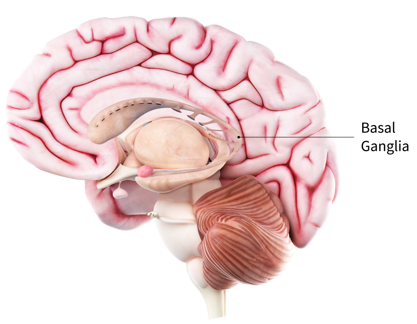
The most prominent signs and symptoms of Parkinson’s disease occur when nerve cells in the basal ganglia (Figure 13.1) , an area of the brain that controls movement, become impaired and/or die. Normally, these nerve cells, or neurons, produce an important brain chemical known as dopamine. When the neurons die or become impaired, they produce less dopamine, which causes the movement problems associated with the disease. Scientists still do not know what causes the neurons to die.
People with Parkinson’s disease also lose the nerve endings that produce norepinephrine, the main chemical messenger of the sympathetic nervous system, which controls many functions of the body, such as heart rate and blood pressure. The loss of norepinephrine might help explain some of the non-movement features of Parkinson’s, such as fatigue, irregular blood pressure, decreased movement of food through the digestive tract, and sudden drop in blood pressure when a person stands up from a sitting or lying position.
Some cases of Parkinson’s disease appear to be hereditary and a few cases can be traced to specific genetic mutations. While genetics is thought to play a role in Parkinson’s, in most cases the disease does not seem to run in families. Many researchers now believe that Parkinson’s results from a combination of genetic and environmental factors, such as exposure to toxins.
Learning Objectives
Upon completion of the work in this chapter students should be able to:
- Describe the composition of gray and white matter and provide examples of brain structures made of each.
- Describe and identify the brain meninges: dura mater, arachnoid mater, & pia mater.
- Define the following structural features of the brain: gyrus, sulcus, fissure.
- Identify brain structures on a dissected brain specimen, model, or diagram.
- Identify cranial nerves on a model or diagram and describe functions of each.
- Identify and define anatomical features of the spinal cord on a model or diagram for both longitudinal view and cross-sectional views.
Background.
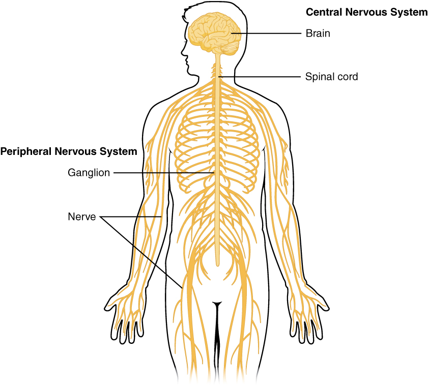
The nervous system can be divided into two major regions: the central and peripheral nervous systems. The central nervous system (CNS) is the brain and spinal cord, and the peripheral nervous system (PNS) is everything else (Figure 13.2). The brain is contained within the cranial cavity of the skull, and the spinal cord is contained within the vertebral cavity of the vertebral column. It is a bit of an oversimplification to say that the CNS is what is inside these two cavities and the peripheral nervous system is outside of them, but that is one way to start to think about it. There are some elements of the peripheral nervous system that are within the cranial or vertebral cavities. The peripheral nervous system is so named because it is on the periphery—meaning beyond the brain and spinal cord. Depending on different aspects of the nervous system, the dividing line between central and peripheral is not necessarily universal.
Nervous Tissue Structures
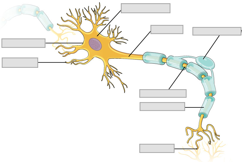
Nervous tissue, present in both the CNS and PNS, contains two basic types of cells: neurons and glial cells. A glial cell is one of a variety of cells that provide a framework of tissue that supports the neurons and their activities and will not be considered further in this lab. The neuron is the more functionally important of the two, in terms of the communicative function of the nervous system.
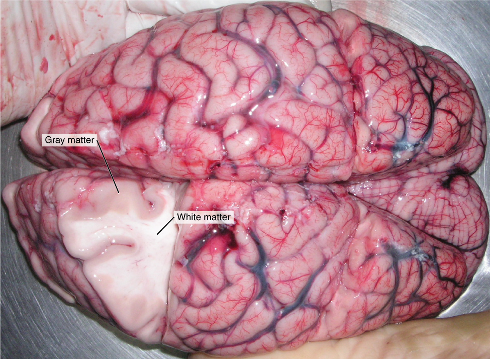
Neurons are cells and therefore have a soma, or cell body, but they also have notable extensions of the cell; each extension is generally referred to as a process. There is one important process that nearly all neurons have called an axon, which is the fiber that connects a neuron with its target. Another type of process that branches off from the soma is the dendrite. Dendrites are responsible for receiving most of the input from other neurons. Looking at nervous tissue, there are regions that predominantly contain cell bodies and regions that are largely composed of axons. These two regions within nervous system structures are referred to as gray matter (the regions with many cell bodies and dendrites) or white matter (the regions with many axons). The colors ascribed to these regions are what would be seen in unstained, nervous tissue (Figure 13.3). Gray matter is not necessarily gray. It can be pinkish because of blood content, or even slightly tan, depending on how long the tissue has been preserved. White matter is white because axons are insulated by a lipid-rich substance called myelin. Lipids can appear as white material, much like the fat on a raw piece of meat. Gray matter may have that color ascribed to it because next to the white matter, it is just darker— hence, gray.
The cell bodies of neurons or axons are often located in discrete anatomical structures that are named. Those names are specific to whether the structure is central or peripheral. A localized collection of neuron cell bodies in the CNS is referred to as a nucleus. In the PNS, a cluster of neuron cell bodies is referred to as a ganglion. A notable exception to this naming convention is a group of nuclei in the central nervous system that were once called the basal ganglia before “ganglion” became accepted as a description for a peripheral structure. Some sources refer to this group of nuclei as the “basal nuclei” which helps avoid confusion.
Terminology applied to bundles of axons also differs depending on location. A bundle of axons, or fibers, found in the CNS is called a tract whereas the same thing in the PNS would be called a nerve. Please note that both can be used to refer to the same bundle of axons. When those axons are in the PNS, the term is nerve, but if they are in the CNS, the term is tract. One example of this is the axons that project from the nervous tissue in the retina into the brain. Axons leaving the eye are called the optic nerve but as soon as they enter the cranium they are referred to as the optic tract.
The Meninges
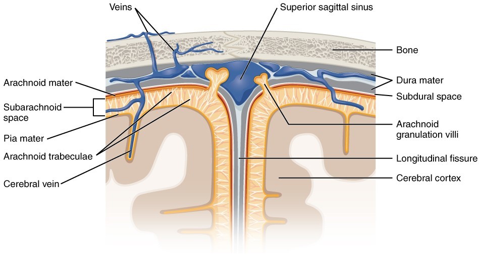
The outer surface of the brain is covered by a series of membranes composed of connective tissue called the meninges, which protect the brain (Figure 13.4). There are three major meningeal layers; the dura mater, the arachnoid mater and the pia mater.
Dura mater
Like a thick cap covering the brain, the dura mater is a tough outer covering. It is a thick fibrous layer and a strong protective sheath over the entire brain and spinal cord. It is anchored to the inner surface of the cranium and to the very end of the vertebral cavity. The name comes from the Latin for “tough mother” to represent its physically protective role. It encloses the entire CNS and the major blood vessels that enter the cranium and vertebral cavity.
Arachnoid mater
The middle layer of the meninges is the arachnoid, named for the spider-web–like extensions between it and the pia mater. The arachnoid defines a sac-like enclosure around the CNS. The branching extensions are found in the subarachnoid space, which is filled with circulating CSF (cerebrospinal fluid). The arachnoid emerges into the dural sinuses as the arachnoid granulations, where the CSF is filtered back into the blood for drainage from the nervous system. The subarachnoid space is filled with circulating CSF, which also provides a liquid cushion to the brain and spinal cord. Like clinical blood work, a sample of CSF can be withdrawn to find chemical evidence of neuropathology or metabolic traces of the biochemical functions of nervous tissue.
Pia mater
Directly adjacent to the surface of the CNS is the pia mater, a thin fibrous membrane that extends into every convolution of gyri and sulci in the cerebral cortex (contours of the brain) and other grooves and indentations. It is thought to have a continuous layer of cells providing a fluid-impermeable membrane. The name pia mater comes from the Latin for “tender mother,” suggesting the thin membrane is a gentle covering for the brain.
Brain Anatomy
The brain and the spinal cord make up the central nervous system, and they represent the main organs of the nervous system. While the spinal cord is a single structure, the adult brain is described in terms of four major regions: the cerebrum, the diencephalon, the brain stem, and the cerebellum.
Cerebrum
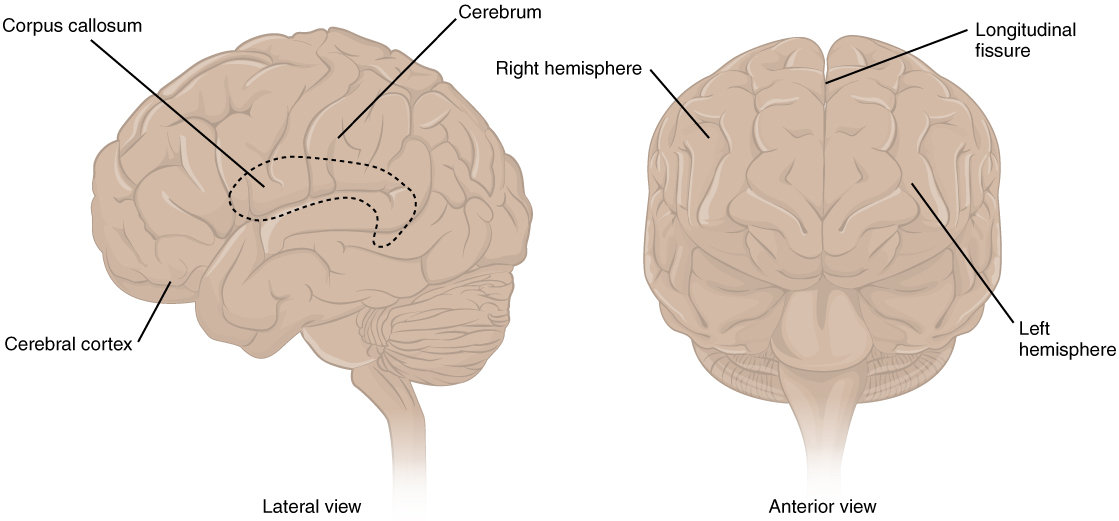
The iconic gray mantle of the human brain, which appears to make up most of the mass of the brain, is the cerebrum (Figure 13.5) The wrinkled outer portion is the cerebral cortex, and the rest of the structure is beneath that outer covering. There is a large separation between the two sides of the cerebrum called
the longitudinal fissure which separates the cerebrum into two distinct halves, a right and left cerebral hemisphere. Deep within the cerebrum, the white matter of the corpus callosum provides the major pathway for communication between the two hemispheres of the cerebral cortex. Many of the higher neurological functions, such as memory, emotion, and consciousness, are the result of cerebral function.
Cerebral cortex
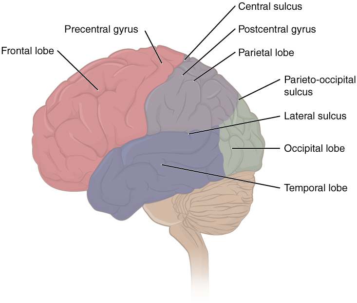
The cerebrum is covered by a continuous layer of gray matter that wraps around either side of the forebrain— the cerebral cortex. This thin, extensive region of wrinkled gray matter is responsible for the higher functions of the nervous system. A gyrus (plural = gyri) is the ridge of one of those wrinkles, and a sulcus (plural = sulci) is the groove between two gyri. The pattern of these folds of tissue can be used to indicate specific regions of the cerebral cortex.
The folding of the cortex maximizes the amount of gray matter in the cranial cavity. During embryonic development, the telencephalon is a structure that eventually develops into the cerebrum. As the telencephalon expands within the skull, the brain goes through a regular course of growth that results in everyone’s brain having a similar pattern of folds. The surface of the brain can be mapped based on the locations of large gyri and sulci. Using these landmarks, the surface of the cortex can be separated into four major regions, or lobes (Figure 13.6). The lateral sulcus that separates the temporal lobe from the other regions is one such landmark. Superior to the lateral sulcus are the parietal and frontal lobes, which are separated from each other by the central sulcus. The posterior region of the cortex is the occipital lobe, which has no obvious anatomical border between it and the parietal or temporal lobes on the lateral surface of the brain. From the medial surface, an obvious landmark separating the parietal and occipital lobes is called the parieto-occipital sulcus. The fact that there is no obvious anatomical border between these lobes is consistent with the functions of these regions being interrelated.
The frontal lobe is responsible for complex functions including motor functions (planning and executing movements via commands sent to the spinal cord and periphery) and, within the prefrontal cortex, aspects of personality via influencing motor responses involved in decision-making. The other lobes are responsible for sensory functions. The parietal lobe is where somatosensation is processed. The occipital lobe is where visual processing begins, although the other parts of the brain can contribute to visual function. The temporal lobe contains the cortical area for auditory processing and also has regions crucial for memory formation.
Located deep within the lateral sulcus is a fifth lobe of the brain called the insular lobe. The function of the insular lobe is not very well understood, however, evidence suggests that it is involved in several processes like motor-control, homeostasis and self awareness. It has also been linked to addiction and a variety of neuropsychiatric disorders.
Subcortical gray matter
Beneath the cerebral cortex are sets of nuclei known as subcortical nuclei that augment cortical processes. The nuclei of the basal forebrain modulate the overall activity of the cortex, possibly leading to greater attention to sensory stimuli. The hippocampus and amygdala are medial-lobe structures that, along with the adjacent cortex, are involved in long-term memory formation and emotional responses.
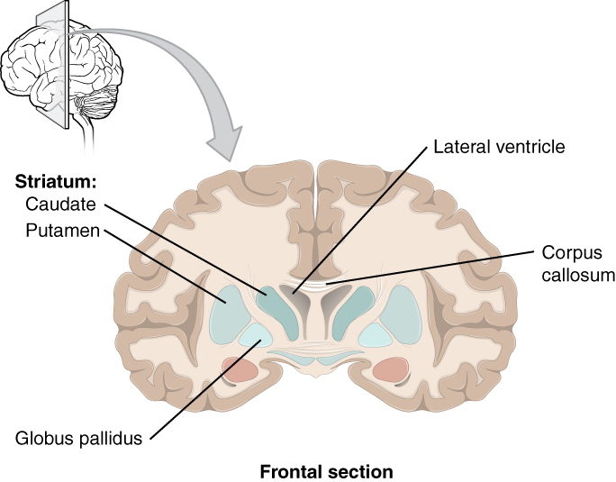
The basal nuclei are a set of nuclei in the cerebrum responsible for comparing cortical processing with the general state of activity in the nervous system to influence the likelihood of movement taking place. The major structures of the basal nuclei that control movement are the caudate, putamen, and globus pallidus, which are located deep in the cerebrum. The caudate is a long nucleus that follows the basic C-shape of the cerebrum from the frontal lobe, through the parietal and occipital lobes, into the temporal lobe. The putamen is mostly deep in the anterior regions of the frontal and parietal lobes. Together, the caudate and putamen are called the striatum. The globus pallidus is a layered nucleus that lies just medial to the putamen; they are called the lenticular nuclei because they look like curved pieces fitting together like lenses. The globus pallidus has two subdivisions, the external and internal segments, which are lateral and medial, respectively. These nuclei can be seen via a frontal section of the brain (Figure 13.7).
Diencephalon
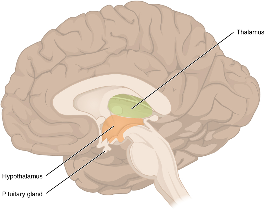
The diencephalon is the connection between the cerebrum and the nearly all of the nervous system and has two major regions: the thalamus and the hypothalamus (Figure 13.8). Most of the brain, the spinal cord, and the PNS send information to the cerebrum through the diencephalon. Output from the cerebrum passes back through the diencephalon to the periphery. The single exception is the system associated with olfaction, or the sense of smell, which connects directly with the cerebrum.
The thalamus is a collection of nuclei that relay information between the cerebral cortex and the periphery, spinal cord, or brain stem. All sensory information, except for olfaction, passes through the thalamus before processing by the cortex. The thalamus does not just pass the information on, it also processes that information. The cerebrum and basal nuclei also send motor information to the thalamus which usually involves interactions between the cerebellum and other nuclei in the brain stem as well.
Inferior and slightly anterior to the thalamus is the hypothalamus, the other major region of the diencephalon. The hypothalamus is a collection of nuclei that are largely involved in regulating homeostasis. The hypothalamus is the executive region in charge of the autonomic nervous system and the endocrine system through its regulation of the anterior pituitary gland. Other parts of the hypothalamus are involved in memory and emotion as part of the limbic system.
Brain stem
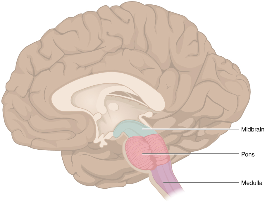
The midbrain and hindbrain (composed of the pons and the medulla) are collectively referred to as the brain stem (Figure 13.9). The structure emerges from the ventral surface of the forebrain as a tapering cone that connects the brain to the spinal cord. Attached to the brain stem, but considered a separate region of the adult brain, is the cerebellum. The midbrain coordinates sensory representations of the visual, auditory, and somatosensory perceptual information. The pons is the main connection with the cerebellum. The pons and the medulla regulate several crucial functions, including the cardiovascular and respiratory systems and rates. The cranial nerves (described below) connect through the brain stem and provide the brain with the sensory input and motor output associated with the head and neck, including most of the special senses. The major ascending and descending pathways between the spinal cord and brain, specifically the cerebrum, pass through the brain stem.
The midbrain is a small region between the thalamus and pons. The upper portion of the midbrain is composed of four bumps known as the colliculi (singular = colliculus), which means “little hill” in Latin.
The inferior colliculus is the inferior pair of these enlargements and is part of the auditory brain stem pathway. Neurons of the inferior colliculus project to the thalamus, which then sends auditory information to the cerebrum for the conscious perception of sound. The superior colliculus is the superior pair and combines sensory information about visual space, auditory space, and somatosensory space. Activity in the superior colliculus is related to orienting the eyes to a sound or touch stimulus.
Cerebellum
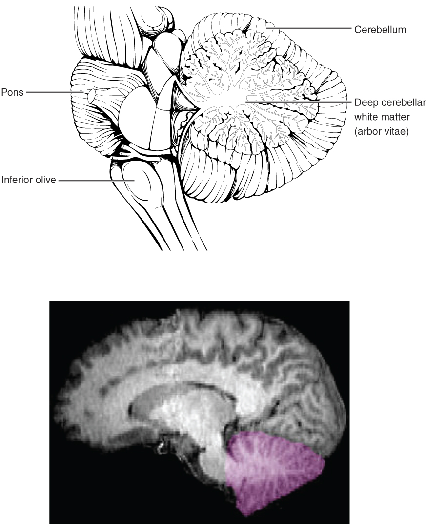
The cerebellum, as the name suggests, is the “little brain” and accounts for approximately 10 percent of the mass of the brain. It is covered in gyri and sulci like the cerebrum, and looks like a miniature version of that part of the brain (Figure 13.10). The cerebellum is largely responsible for comparing information from the cerebrum with sensory feedback from the periphery through the spinal cord.
Descending fibers from the cerebrum have branches that connect to neurons in the pons. Those neurons project into the cerebellum, providing the cerebellum with the same motor information that is sent to the spinal cord. Sensory information from the periphery, which enters through spinal or cranial nerves, also projects to a nucleus in the medulla known as the inferior olive. Fibers from this nucleus enter the cerebellum and are compared with the descending commands from the cerebrum. For example, if the cerebrum sends a command down to the spinal cord to initiate walking, a copy of that motor command is sent to the cerebellum. Sensory feedback from the muscles and joints, proprioceptive information about the movements of walking, and sensations of balance are sent to the cerebellum through the inferior olive and then the cerebellum integrates all of that information. If walking is not coordinated, perhaps because the ground is uneven or a strong wind is blowing, then the cerebellum sends out a corrective command to compensate for the difference between the original command from the cerebrum and the sensory feedback from the periphery. The output of the cerebellum is into the midbrain, which then sends a descending input to the spinal cord to correct motor information going to skeletal muscles.
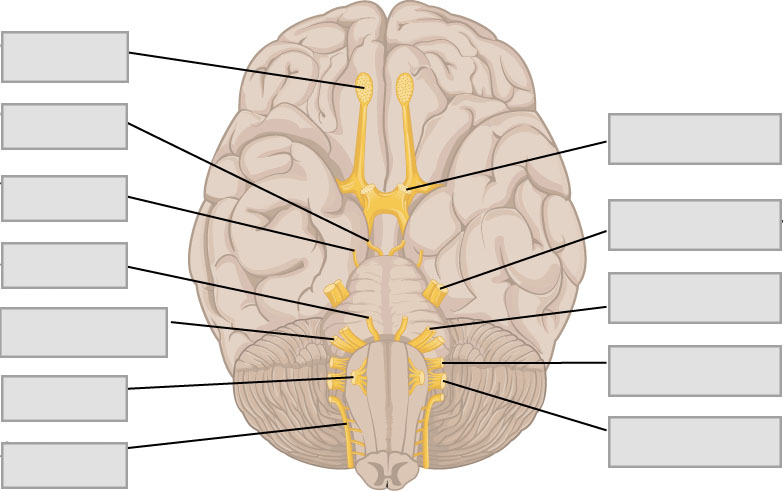
Cranial Nerves
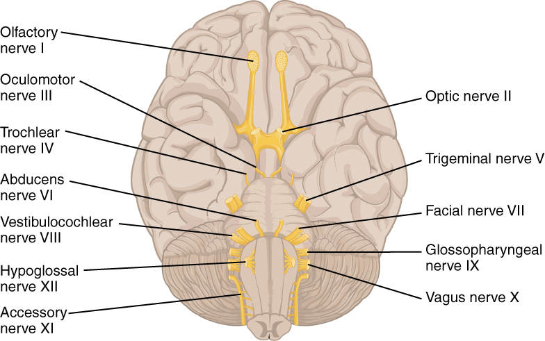
The nerves attached to the brain are the cranial nerves, which are primarily responsible for the sensory and motor functions of the head and neck (one of these nerves targets organs in the thoracic and abdominal cavities as part of the parasympathetic nervous system) (Figure 13.11, Table 13.1). There are twelve cranial nerves, which are designated CNI through CNXII for “Cranial Nerve,” using Roman numerals for 1 through 12.
The olfactory nerve (I) and optic nerve (II) are responsible for the sense of smell and vision, respectively. The oculomotor nerve (III) is responsible for eye movements by controlling four of the extraocular muscles. It is also responsible for lifting the upper eyelid when the eyes point up, and for pupillary constriction. The trochlear nerve (IV) and the abducens nerve (VI) are both responsible for eye movement but do so by controlling different extraocular muscles. The trigeminal nerve (V) is responsible for cutaneous sensations of the face and controlling the muscles of mastication. The facial nerve (VII) is responsible for the muscles involved in facial expressions, as well as part of the sense of taste and the production of saliva.
The vestibulocochlear nerve (VIII) is responsible for the senses of hearing and balance. The glossopharyngeal nerve (IX) is responsible for controlling muscles in the oral cavity and upper throat, as well as part of the sense of taste and the production of saliva. The vagus (X) nerve is responsible for contributing to homeostatic control of the organs of the thoracic and upper abdominal cavities via autonomic neurons. The spinal accessory nerve (XI) is responsible for controlling the muscles of the neck, along with cervical spinal nerves. The hypoglossal nerve (XII) is responsible for controlling the muscles of the lower throat and tongue.
The cranial nerves can be classified as sensory nerves, motor nerves, or a combination of both, meaning that the axons in these nerves can originate out of sensory ganglia external to the cranium or motor nuclei within the brain stem. Sensory axons enter the brain to synapse in a nucleus. Motor axons connect to skeletal muscles of the head or neck. Three of the nerves are solely composed of sensory fibers; five are strictly motor; and the remaining four are mixed nerves that contain both sensory and motor fibers. The first, second, and eighth nerves are purely sensory (olfactory (CNI), optic (CNII), and vestibulocochlear (CNVIII) nerves). The three eye-movement nerves are all motor (oculomotor (CNIII), trochlear (CNIV), and abducens (CNVI)). The spinal accessory (CNXI) and hypoglossal (CNXII) nerves are also strictly motor. The remainder of the nerves (trigeminal (CNV), facial (CNVII), glossopharyngeal (CNIX), and vagus (CNX) nerves) contain both sensory and motor fibers and are often related to each other. The trigeminal and facial nerves both concern the face; one is primarily associated the sensations and the other primarily associated with the muscle movements. The facial and glossopharyngeal nerves are both responsible for conveying gustatory, or taste, sensations as well as controlling salivary glands. The vagus nerve is involved in visceral responses to taste, namely the gag reflex.
An important learning outcome for this lesson is to understand and describe the functions of cranial nerves. While this can feel a lot of information to commit to memory, it is possible by using memory tools like mnemonics. There are many mnemonics others have created that can quickly be found via an internet search. However, the best way to remember a mnemonic, is to make your own with personally-relatable information (i.e. movies, sports, friends names, etc.).
| Table 13.1 The Cranial Nerves
|
|||
| Number | Name | Type | Function(s) |
| I | Olfactory | Sensory | • Sensory information from the nose. |
| II | Optic | Sensory | • Sensory information from the eyes. |
| III | Oculomotor | Motor | • Motor information to most rectus and inferior oblique muscles to cause eye movement. |
| IV | Trochlear | Motor | • Motor information to superior oblique muscle for eye movement. |
| V | Trigeminal | Both | • Sensory information from and motor information to the face.
• Motor information for chewing. |
| VI | Abducens | Motor | • Motor information to lateral rectus muscle to cause eye movement. |
| VII | Facial | Both | • Sensory information from anterior part of the tongue.
• Motor information to the face. • Innervates lacrimal, salivary and other glands. |
| VIII | Vestibulocochlear | Sensory | • Sensory information from the ear for hearing and equilibrium. |
| IX | Glossopharyngeal | Both | • Sensory information from posterior part of the tongue.
• Motor information to tongue and throat. |
| X | Vagus | Both | • Sensory information from abdomen, thorax, neck and root of tongue.
• Motor information to heart, digestive organs, spleen and kidneys. |
| XI | Accessory | Motor | • Motor information for swallowing. |
| XII | Hypoglossal | Motor | •Motor information to the tongue. |
The Spinal Cord
Anatomy of Spinal Cord
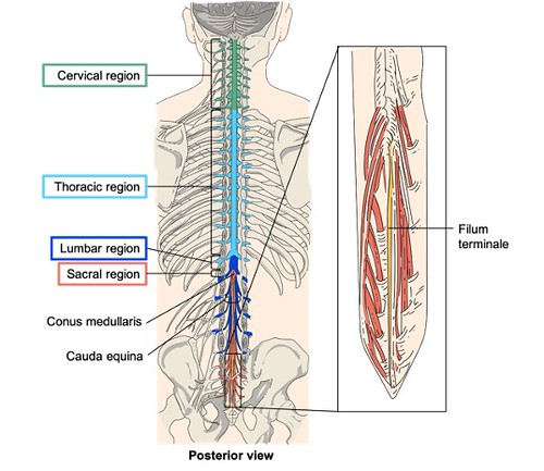
In an adult, the spinal cord is about eighteen inches long and extends from the foramen magnum of the skull to approximately the first lumbar vertebra and is divided into regions that correspond to regions of the vertebral column (Figure 13.12). The name of each spinal cord region corresponds to the level at which spinal nerves pass through the intervertebral foramina. Immediately adjacent to the brain stem is the cervical region, followed by the thoracic, then the lumbar, and finally the sacral region. The spinal cord has two areas where the diameter of the spinal cord is enlarged because of increased neural structures associated with the appendages. The cervical enlargement is caused by nerves moving to and from the arms and is located from approximately C3 through T2. The lumbar enlargement is caused by nerves moving to and from the legs and is located from about T7 through T11 (Figure 13.12).
The spinal cord does not extend the full length of the vertebral column because the spinal cord does not grow significantly longer after the first or second year while the skeleton continues to grow. As the vertebral column continues to grow, spinal nerves grow with it and result in a long bundle of nerves that resembles a horse’s tail, called the cauda equina (Figure 13.12). Some of the largest neurons of the spinal cord extend from the cauda equina including the motor neuron that causes contraction of the big toe which is located in the sacral region of the spinal cord. This motor neuron’s axon reaches all the way to the belly of that muscle which can be over a meter in distance in a tall person. The neuronal cell body that maintains that long fiber is also necessarily quite large, possibly several hundred micrometers in diameter, making it one of the largest cells in the body. Immediately superior to the cauda equina, the spinal cord terminates at the medullary cone (also known as the conus medullaris) at approximately vertebra L1. Beyond the medullary cone, the meninges that cover the spinal cord (discussed below) continue as a thin, delicate strand of tissue called the terminal filum, which anchors the spinal cord to the coccyx.
31 pairs of spinal nerves extend from the spinal cord and each pair is named for the level of the spinal cord from which each pair emerges. There are eight pairs of cervical nerves designated C1 to C8, twelve thoracic nerves designated T1 to T12, five pairs of lumbar nerves designated L1 to L5, five pairs of sacral nerves designated S1 to S5, and one pair of coccygeal nerves. The first nerve, C1, emerges between the first cervical vertebra and the occipital bone. The second nerve, C2, emerges between the first and second cervical vertebrae. The same occurs for C3 to C7, but C8 emerges between the seventh cervical vertebra and the first thoracic vertebra. For the thoracic and lumbar nerves, each one emerges between the vertebra that has the same designation and the next vertebra in the column. The sacral nerves emerge from the sacral foramina along the length of that unique vertebra.
The Meninges (of the Spinal Cord)
The spinal cord and brain are covered by the meninges which are a continuous, layered unit of tissues that provide support and protection to the delicate structures of the nervous system. The meninges include three layers: the dura mater, arachnoid mater, and pia mater (Figure 13.4). The outermost layer, the dura mater, is anchored to the inside of the vertebral cavity. It is thick and “dura”ble, providing protection and support to the spinal cord. The arachnoid mater is the thin middle layer, connecting the dura mater to the pia mater. The arachnoid mater gets its name from its web-like appearance and is connected to the pia mater through tiny fibrous extensions that span the subarachnoid space between the two layers. The innermost pia mater is in direct contact with the spinal cord and brain. It is thin and rich in blood vessels, although the pia mater is thicker and less vascular in the spinal cord than in the brain.
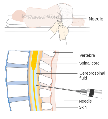
The subarachnoid space is filled with cerebrospinal fluid (CSF) which protects the CNS by providing cushioning. In order to test for disease or dysfunction in the central nervous system, CSF may be removed and analyzed via a procedure called a spinal tap or lumbar puncture (Figure 13.13). Because of the close proximity between the meninges and nervous tissue, this procedure is typically done at the end of the spinal cord, where the terminal filum extends from the inferior end of CNS at the upper lumbar region to the sacral end of the vertebral column. Because the spinal cord does not extend through the lower lumbar region of the vertebral column, a needle can be inserted in this region through the dura and arachnoid layers to withdraw CSF with minimal risk of damaging the nervous tissue of the spinal cord. One example of a disease commonly diagnosed via lumbar puncture is meningitis, which is an inflammation of the meninges caused by either a viral or bacterial infection. Symptoms include fever, chills, nausea, vomiting, sensitivity to light, soreness of the neck, and severe headache. More serious are the possible neurological symptoms, such as changes in mental state including confusion, memory deficits, other dementia-type symptoms, hearing loss, and even death due to the close proximity of the infection to nervous system structures.
Cross-sectional Anatomy of the Spinal Cord
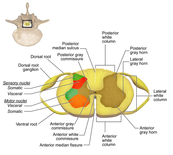
Each section of the spinal cord has its associated spinal nerves forming two nerve routes that include a combination of incoming sensory axons and outgoing motor axons. For example, the radial nerve contains fibers of cutaneous sensation in the arm, as well as motor fibers that move muscles in the arm. The sensory axons that form a part of the radial nerve enter the spinal cord as the posterior (dorsal) nerve root, whereas the motor fibers emerge as the anterior (ventral) nerve root (Figure 13.14). The cell bodies of sensory neurons are grouped together at the posterior (dorsal) root ganglion, causing an enlargement of that portion of the spinal nerve. Note that it is common to see the terms dorsal and ventral used interchangeably with posterior and anterior, particularly in reference to nerves and the structures of the spinal cord.
Inside the spinal cord, the anterior and posterior nerve roots form the gray matter of the spinal cord. In cross section (Figure 13.14), the distribution of gray matter of the spinal cord is often compared to an inkblot test or butterfly, with the spread of the gray matter, subdivided into regions referred to as horns, on one side replicated on the other. The posterior horn receives information from the posterior nerve root and is therefore responsible for sensory processing, while the anterior horn sends out motor signals to the anterior nerve root to move skeletal muscles. The lateral horn, which is only found in the thoracic, upper lumbar, and sacral regions, is a key component of the sympathetic division of the autonomic nervous system. The anterior median fissure marks the anterior midline and the posterior median sulcus marks the posterior midline. Each side of the gray matter is connected by the gray commissure and located in the center of the gray commissure is the central canal, which runs the length of the spinal cord. The central canal is continuous with the ventricular system of the brain and transports nutrients to the spinal cord.
Comparable to the gray matter being separated into horns, the white matter of the spinal cord is separated into columns. Ascending tracts of nervous system fibers in these columns carry sensory information from the periphery to the brain, whereas descending tracts carry motor commands from the brain to the periphery. Looking at the spinal cord longitudinally, the columns extend along its length as continuous bands of white matter. In cross-section, the posterior columns can be seen between the two posterior horns of gray matter, whereas the anterior columns are bounded by the anterior horns. The white matter on either side of the spinal cord, between the posterior horn and the anterior horn, are the lateral columns. The posterior columns are composed of axons of ascending tracts, whereas the anterior and lateral columns are composed of many different groups of axons of both ascending and descending tracts.
Spinal Nerve Plexuses
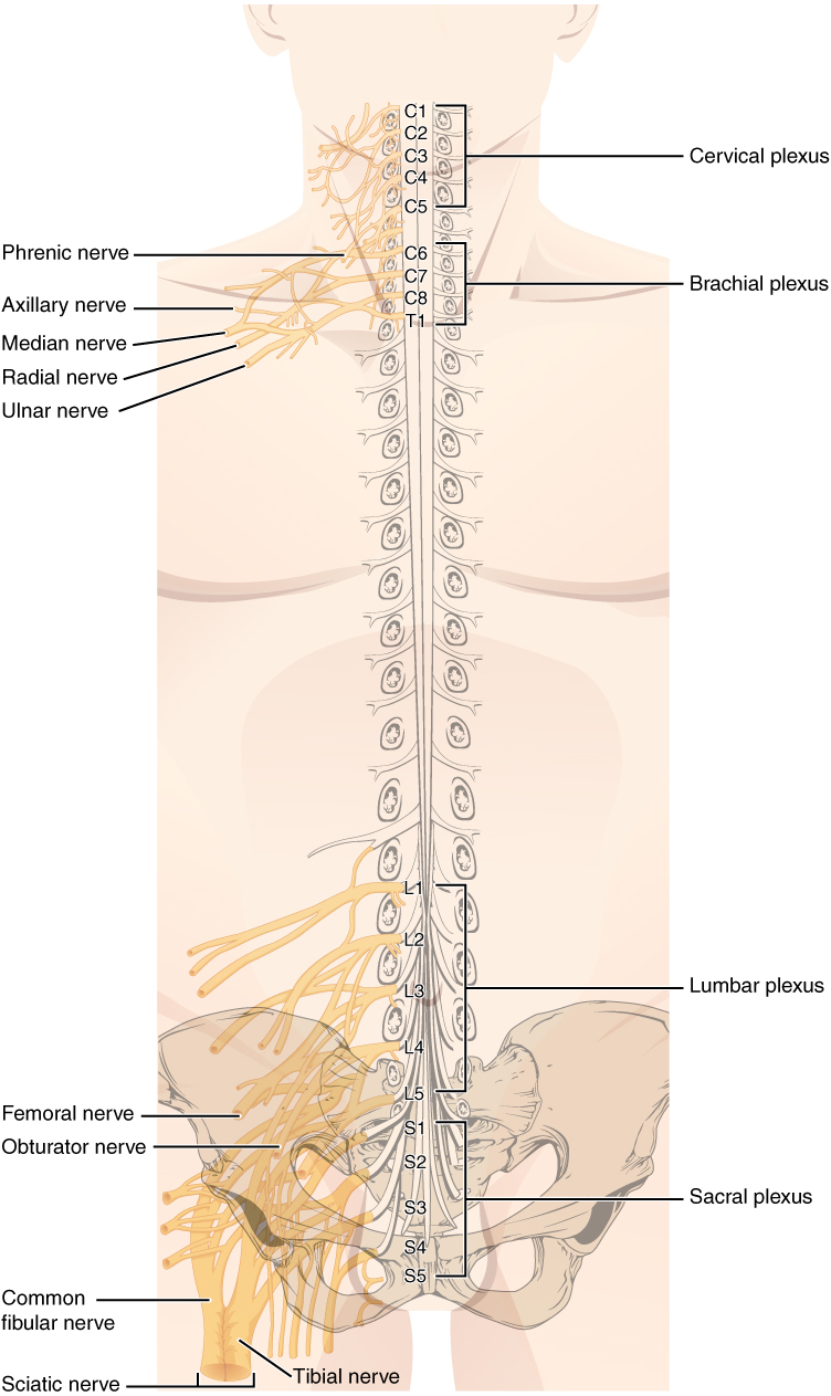
Spinal nerves extend outward from the vertebral column to innervate the periphery. The nerves in the periphery are not straight continuations of the spinal nerves, but rather the reorganization of the axons in those nerves to follow different courses. Axons from different spinal nerves will come together to form a peripheral nerve. This occurs at four places along the length of the vertebral column, each identified as a nerve plexus which have previously been described in the context of the peripheral nerves. Focusing on the relationship to spinal nerves, two nerve plexuses are found at the cervical level, one at the lumbar level, and one at the sacral level (Figure 13.15). The cervical plexus is composed of axons from spinal nerves C1 through C5 and branches into nerves in the posterior neck and head, as well as the phrenic nerve, which connects to the diaphragm at the base of the thoracic cavity. The other plexus from the cervical level is the brachial plexus. Spinal nerves C4 through T1 reorganize through this plexus to give rise to the nerves of the arms (ex: radial nerve), as the name brachial suggests. The lumbar plexus arises from all the lumbar spinal nerves and gives rise to nerves innervating the pelvic region and the anterior leg (ex: femoral nerve). The sacral plexus comes from the lower lumbar nerves L4 and L5 and the sacral nerves S1 to S4. The most significant systemic nerve to come from this plexus is the sciatic nerve, which is a combination of the tibial nerve and the fibular nerve. Spinal nerves of the thoracic region, T2 through T11, are not part of the plexuses but rather emerge and give rise to the intercostal nerves, which innervate the intercostal muscles found in between ribs.
| Table 13.2 Nerve plexuses | ||
| Associated Spinal Nerves | Major Associated Peripheral Nerves | |
| Cervical | C1-5 | Phrenic |
| Brachial | C5-T1 | Radial, median, ulnar, musculocutaneous, axillary |
| Lumbar | L1-4 | Femoral, obturator |
| Sacral | L4-S4 | Sciatic |
Pre-Laboratory Questions
- Identify the parts of a typical neuron: Axon, cell body, dendrites , nucleus, axon terminals.
- Which of the following is not a function of the nervous system?
A. stimulate muscles and glands
B. contribute to homeostatic feedback loops
C. produce quick effects by electrochemical mechanisms
D. release chemicals into the bloodstream for distribution throughout the body
3. Which part of the brain has noticeable superior and lateral gyri (folds) that increase surface area for cortical gray matter?
A. midbrain
B. pons
C. cerebellum
D. brainstem
E. left and right hemispheres
4. In addition to meninges, fluid-filled spaces help protect the brain nervous tissue. The subarachnoid space would be found between which two meninges?
A. arachnoid and pia mater
B. dura mater and arachnoid mater
C. dura mater and pia mater
5. Which of the following is not important for creating the Blood-brain barrier (BBB)?
A. tight ependymal cell junctions
B. basement membrane
C. tight endothelial (capillary wall epithelium) cell junctions
D. ependymal cilia
Exercises
- Exercise 1 Gray and white mater composition and brain areas
- Exercise 2 Identification of brain meninges: dura mater, arachnoid mater, & pia mater
- Exercise 3 Identification of brain structures on a dissected brain specimen, model, or diagram
- Exercise 4 Identification of cranial nerves
- Exercise 5 Anatomical features of the spinal cord on a model or diagram
Exercise 1 Gray and white mater composition and brain areas
Required materials
- None
Procedure
- This activity will be completed individually or in small groups. Refer to the background information to answer the question below.
- Define the following terms and provide examples of each in the central nervous system
| Terms | Definition | Examples in CNS |
| Gray mater
|
||
| White mater |
Exercise 2 Identification of brain meninges: dura mater, arachnoid mater, & pia mater
Required materials
- None
Procedure
- This activity will be completed individually or in small groups. Refer to the background information to answer the questions below.
- Categorize the following terms and provide a one line definition for each of them. For the meninges, also rank them from the most superficial layer to the deepest layer.
Gyri, pia mater, sulcus, arachnoid mater, fissure, dura mater
| Brain meninges | Definition |
| Superficial– | |
| Deepest– | |
| Brain structures | Definition |
Exercise 3 Identification of brain structures on a dissected brain specimen, model, or diagram
Required materials
- Gloves
- Dissection tray
- Dissection instruments
- Sheep brain specimen (or cow brain specimen)
- T-pins for labeling
- Labeling tape
- Sheep brain bismount model
- Brain Cavities Model
- Brain Ventricles Model
- Classic Human Brain Model
- Nervous System on Board Model
- Brain poster
- Nervous system poster
Procedure
- The first activity will be completed individually.
- Refer to the background information and label the following diagram with the appropriate structures.
- The brain dissection activity will be completed in groups of 3-4 in the lab. Please read the following steps carefully before you begin.
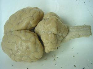
- Place the brain specimen in the tray, dorsal side up (Figure 13.16).
-
- Identify the cerebrum, the longitudinal fissure and the two hemispheres of the brain. You can also locate examples of gyri, sulci and the different lobes of the cerebrum.
-
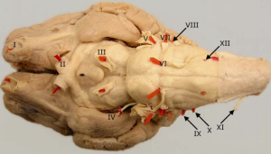
- Place the brain in the tray ventral side up (Figure 13.17) and identify the cerebellum, pons, medulla and optic chiasma.
-
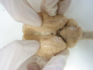
- Place the brain on the tray, dorsal side up. Locate the longitudinal fissure and gently try to widen it with your fingers (Figure 13.18).
-
- Insert a scalpel in the fissure and cut through the brain into two longitudinal halves (Figure 13.19).
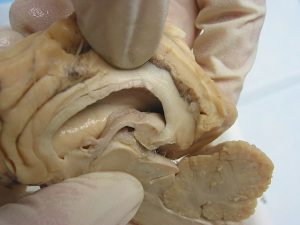
-
- With the cut sides facing up, identify the thalamus, hypothalamus, pineal body, pons and medulla.
- Locate the corpus collosum and lateral ventricles.
- Observe the cut surface of the cerebellum and try to identify the tree like structure made of white mater called arbor vitae or “tree of life”.
- Compare the structures that you see in your dissected samples to those from other groups.
- Your instructor will help you identify the same structures on a dissected human brain.
- Using T-pins and labeling tape, label all the regions of the brain you identified. Take a picture (or pictures) and insert in the space below.

-
- When you are done observing and taking pictures of the sheep brain specimen, dispose it off in the biohazard bin and clean the dissecting tray, T-pins and dissection instruments.
Exercise 4 Identification of cranial nerves
Required materials
- None
Procedure
- This activity will be completed individually. Refer to the background information to answer the questions below.
- For each cranial nerve:
-
- Use the summary table (Table 13.1) as your source for this information.
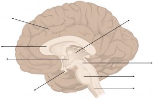
- Identify and label this diagram with appropriate cranial nerves. Identify by both name and number on diagram.
-
- Fill in the blanks to complete the table.
- Identify by both name and number.
- Provide one example of a function.
- Identify whether each nerve carries sensory information, motor information, or both types of information.
- Fill in the blanks to complete the table.
| Name | Number | Type | Function |
| Vestibulocochlear
|
|||
| V
|
|||
| Both | Motor information to the face.
|
||
| Oculomotor
|
|||
| X
|
Exercise 5 Anatomical features of the spinal cord on a model or diagram
Required Materials
- Spinal cord cross section model
- Nervous System on Board model
- The nervous system poster
- Muscular system poster
Procedure
- Identify and define anatomical features of the spinal cord on a model or diagram for both longitudinal view and cross-sectional views.
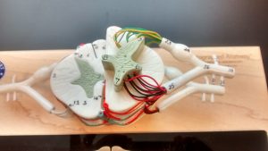
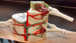
Identify the following features on the spinal cord model. Then draw diagrams (cross section and longitudinal section) to label and show these below.
-
- Posterior (dorsal) median sulcus
- Anterior (ventral) median fissure
- Posterior (dorsal) horn
- Anterior (ventral) horn
- Lateral horn
- Gray commissure
- Posterior (dorsal) root
- Posterior (dorsal) root ganglion
- Anterior (ventral) root
- Posterior (dorsal) column
- Anterior (ventral) column
- Lateral column
- Central canal
- Pia mater
- Arachnoid mater
- Subarachnoid space
- Dura mater
- Spinal nerve

2. Name the layers of the meninges is from superficial to deep:
Post-laboratory Questions
- Which of the following gray or white matter brain structures is correctly paired with its description?
A. projection tracts: receive sensory input and process information locally
B. association tracts: cells with unmyelinated connections within part of the CNS
C. commissural tracts: connects right and left hemisphere
E. neocortex: tracts extending from higher to lower regions of the brain
E. stellate cells: connect different parts within the same hemisphere
2. Which part of the forebrain plays a major role in homeostasis through both the endocrine system and autonomic nervous system?
A. insula
B. corpus callosum
C. hypothalamus
D. precentral gyrus
E. basal nuclei
3. Which part of the cerebral cortex contains the visual center?
A. temporal lobe
B. occipital lobe
C. frontal lobe
D. parietal lobe
E. insula lobe
4. Which of the cranial nerves does not have a sensory component?
A. Facial (VII)
B. Trochlear (IV)
C. Vestibulocochlear (VIII)
D. Olfactory (I)
E. Optic (II)
5. Which of the plexuses would be involved in breathing and movement of the head?
A. coccygeal
B. brachial
C. sacral
D. lumbar
E. cervical
