
7 Chapter 7 Axial Skeleton
By Ganesan L. Kamatchi
Motivation.
Spinal disease refers to a condition impairing the backbone. There are many recognized spinal diseases, some more common than others. Spinal disease also includes cervical spine diseases, which are diseases in the vertebrae of the neck. A lot of flexibility exists within the cervical spine and because of that, it is common for an individual to damage that area, especially over a long period of time. Some of the common cervical spine diseases include degenerative disc disease, cervical stenosis, and cervical disc herniation. Degenerative disc disease occurs over time when the discs within each vertebra in the neck begin to fall apart and begin to disintegrate. Because each vertebra can cause pain in different areas of the body, the pain from the disease can be sensed in the back, leg, neck area, or even the arms. When the spinal canal begins to lose its gap and gets thinner, it can cause pain in the neck, which can also cause a numb feeling in the arms and hands. Those are symptoms of cervical stenosis disease. The discs between each vertebra have fibers that can begin to deteriorate, and this can occur in cervical disc herniation. This disease is less common in younger people as it is usually a function of aging.
Learning Objectives
Upon completion of the work in this chapter students should be able to:
- Identify and label the main bones and structures of the skull from multiple views
- Label and describe the five regions of the vertebral column and vertebrae within
- Identify and describe the attachments and organization of the ribs in the thoracic cage region
Background.
The skeletal system includes all of the bones, cartilages, and ligaments of the body that support and give shape to the body and body structures. The skeleton consists of the bones of the body. For adults, there are 206 bones in the skeleton. Younger individuals have higher numbers of bones because some bones fuse together during childhood and adolescence to form an adult bone. The primary functions of the skeleton are to provide a rigid, internal structure that can support the weight of the body against the force of gravity, and to provide a structure upon which muscles can act to produce movements of the body. The lower portion of the skeleton is specialized for stability during walking or running. In contrast, the upper skeleton has greater mobility and ranges of motion, features that allow you to lift and carry objects or turn your head and trunk.
In addition to providing for support and movements of the body, the skeleton has protective and storage functions. It protects the internal organs, including the brain, spinal cord, heart, lungs, and pelvic organs. The bones of the skeleton serve as the primary storage site for important minerals such as calcium and phosphate. The bone marrow found within bones stores fat and houses the blood-cell producing tissue of the body.
The skeleton is subdivided into two major divisions—the axial and appendicular.
The axial skeleton forms the vertical, central axis of the body and includes all bones of the head, neck, chest, and back (Figure 7.2). It serves to protect the brain, spinal cord, heart, and lungs. It also serves as the attachment site for muscles that move the head, neck, and back, and for muscles that act across the shoulder and hip joints to move their corresponding limbs.

The axial skeleton of the adult consists of 80 bones, including the skull, the vertebral column, and the thoracic cage. The skull is formed by 22 bones. Also associated with the head are an additional seven bones, including the hyoid bone and the ear ossicles (three small bones found in each middle ear). The vertebral column consists of 24 bones, each called a vertebra, plus the sacrum and coccyx. The thoracic cage includes the 12 pairs of ribs, and the sternum, the flattened bone of the anterior chest.
Anterior View of the Skull
The anterior skull consists of the facial bones and provides the bony support for the eyes and structures of the face. This view of the skull is dominated by the openings of the orbits and the nasal cavity. Also seen are the upper and lower jaws, with their respective teeth (Figure 7.3).
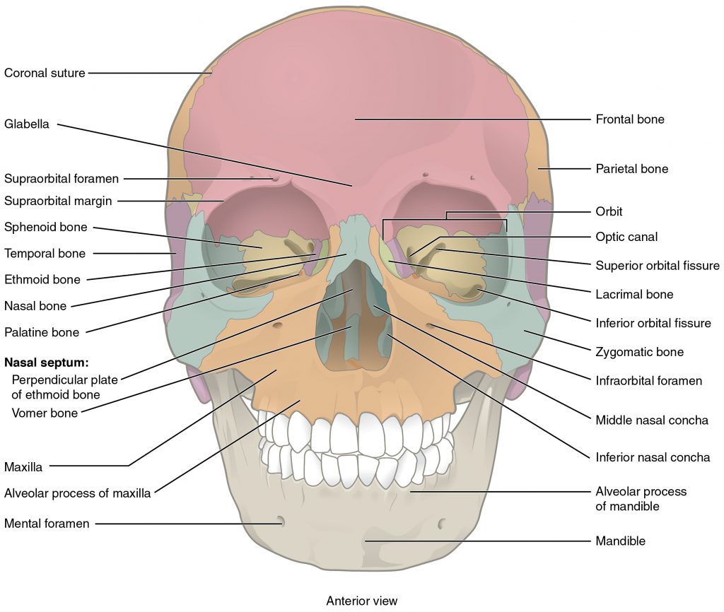
The orbit is the bony socket that houses the eyeball and muscles that move the eyeball or open the upper eyelid. Inside the nasal area of the skull, the nasal cavity is divided into halves by the nasal septum. Each side of the nasal cavity is triangular in shape, with a broad inferior space that narrows superiorly. When looking into the nasal cavity from the front of the skull, three bony plates are seen projecting from each lateral wall, called superior, middle and inferior nasal conchae respectively.
Lateral View of the Skull
A view of the lateral skull is dominated by the large, rounded brain case above and the upper and lower jaws with their teeth below (Figure 7.4). Separating these areas is the bridge of bone called the zygomatic arch. One of the major muscles that pulls the mandible upward during biting and chewing arises from the zygomatic arch. On the lateral side of the brain case, above the level of the zygomatic arch, is a shallow space called the temporal fossa. Below the level of the zygomatic arch and deep to the vertical portion of the mandible is another space called the infratemporal fossa. Both the temporal fossa and infratemporal fossa contain muscles that act on the mandible during chewing.

Posterior View of Skull
Parietal, temporal and occipital bones form the posterior side of the skull. The occipital bone is the single bone that forms the posterior skull and posterior base of the cranial cavity (Figure 7.5). On its outside surface, at the posterior midline, is a small protrusion called the external occipital protuberance, which serves as an attachment site for a ligament of the posterior neck. Lateral to either side of this bump is a superior nuchal line (nuchal = “nape” or “posterior neck”). The nuchal lines represent the most superior point at which muscles of the neck attach to the skull, with only the scalp covering the skull above these lines. On the base of the skull, the occipital bone contains the large opening of the foramen magnum, which allows for passage of the spinal cord as it exits the skull. On either side of the foramen magnum is an oval-shaped occipital condyle. These condyles form joints with the first cervical vertebra and thus support the skull on top of the vertebral column.

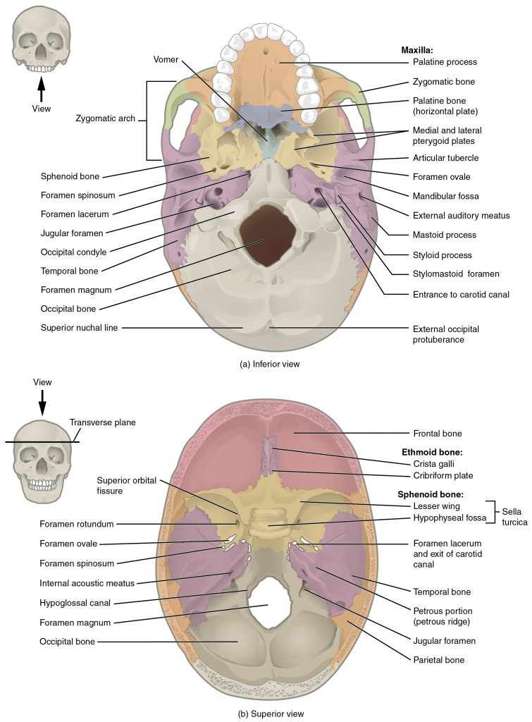
Superior View of Cranial Floor
The base of skull, also known as the cranial base or the cranial floor, is the most inferior area of the skull. It is composed of the endocranium and the lower parts of the skull roof. There are three depressions called cranial fossa is formed by the floor of the cranial cavity. They are anterior cranial fossa, housing the projecting frontal lobes of the brain; middle cranial fossa, separated from the posterior fossa by the clivus and the petrous crest and the posterior cranial fossa, between the foramen magnum and tentorium cerebelli, containing the brainstem and cerebellum (Figure 7.6).
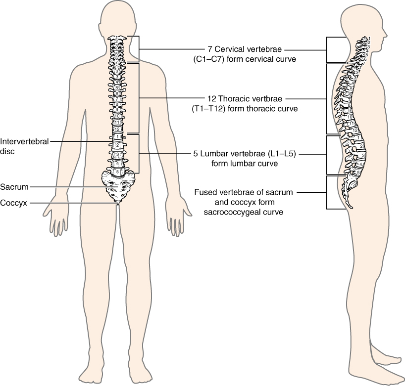
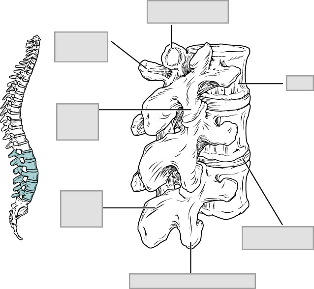
Vertebral Column
The adult vertebral column consists of 24 vertebrae, plus the sacrum (5 vertebrae) and coccyx (4 vertebrae). The vertebrae are divided into five regions: cervical C1–C7 vertebrae, thoracic T1–T12 vertebrae, lumbar L1–L5 vertebrae, sacral S1-S5 vertebrae (fused) and coccygeal Co1-Co4 vertebrae (fused). The vertebral column is curved, with two primary curvatures (thoracic and sacrococcygeal curves) and two secondary curvatures (cervical and lumbar curves). (Figure 7.7). A typical vertebra is shown in Figure 7.8.
Vertebra

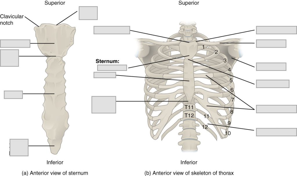
The Thoracic Cage
The thoracic cage is formed by the sternum and 12 pairs of ribs with their costal cartilages. The ribs are anchored posteriorly to the 12 thoracic vertebrae. The sternum consists of the manubrium, body, and xiphoid process. The ribs are classified as true ribs (1–7) and false ribs (8–12). The last two pairs of false ribs are also known as floating ribs (11–12). (Figure 7.9)

Pre-Laboratory Questions
After reviewing the background information, please answer these pre-laboratory questions.
- What are the components of the axial skeleton?
- List the names of the flat bones of the skull.
- Name the five regions of the vertebral column.
- What are the three types of ribs based on their attachments?
Exercises
Type your exercises here.
- Exercise 1 Anterior View of the Skull
- Exercise 2 Lateral View of the Skull
- Exercise 3 Posterior View of Skull
- Exercise 4 Superior View of Cranial Floor
- Exercise 5 Vertebral Column
- Exercise 6 Thoracic Cage
Exercise 1 Anterior View of the Skull
Required Materials
- Skull models
- Poster of the skeletal system
Procedure
- Obtain a skull model and observe its structures from the anterior side (Figure 7.3)
- Identify the structures listed below on the skull and label the structures on the image below.
· Alveolar processes · Optic canal · Frontal bone · Parietal bone · Inferior nasal concha · Perpendicular plate of ethmoid · Inferior orbital fissure · Sphenoid bone · Infraorbital foramen · Superior orbital fissure · Lacrimal bone · Supraorbital foramen · Mandible · Supraorbital margin · Maxilla · Temporal bone · Mental protuberance · Vomer · Nasal bone · Zygomatic bone 
Exercise 2 Lateral View of the Skull
Required Materials
- Skull models
- Poster of the skeletal system
Procedure
- Obtain a skull model and observe its structures from the lateral side (Figure 7.4)
- Identify the structures listed below on the skull and label the structures on the image below.
· Coronal suture · Mental protuberance · Ethmoid bone · Nasal bone · External acoustic meatus · Occipital bone · Frontal bone · Parietal bone · Greater wing of sphenoid bone · Pterion · Lacrimal bone · Squamous part of temporal · Lacrimal fossa · Squamous suture · Lambdoid suture · Styloid process · Mandible · Temporal process · Mastoid process · Zygomatic bone · Maxilla · Zygomatic process 
Exercise 3 Posterior View of Skull
Required Materials
- Skull models
- Poster of the skeletal system
Procedure
- Obtain a skull model and observe its structures from the posterior side (Figure 7.5)
- Identify the structures listed below on the skull and label the structures on the image below.
· External occipital protuberance · Parietal bone · Foramen magnum · Sagittal suture · Lambdoid suture · Superior nuchal line · Mastoid process · Temporal bone · Occipital bone · Zygomatic bone · Occipital condyle 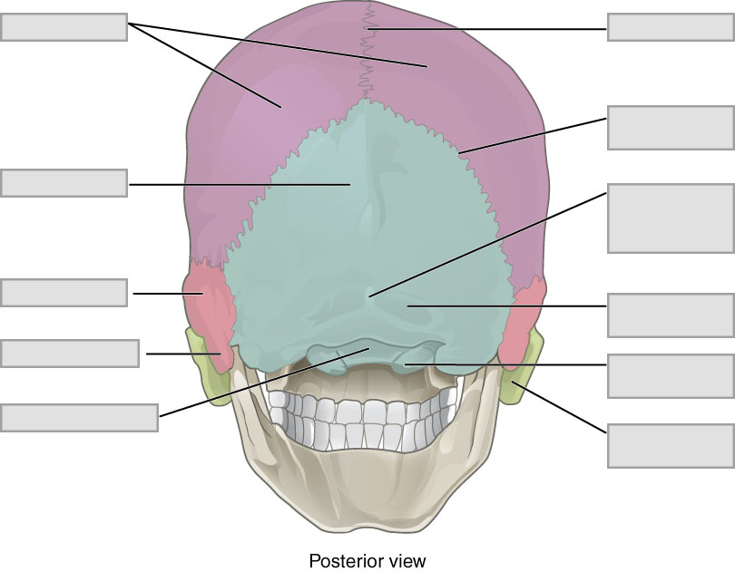
Exercise 4 Superior View of Cranial Floor
Required Materials
- Skull models
- Poster of the skeletal system
Procedure
- Obtain a skull cut along its transverse plane and observe its structures from the cranial floor (Figure 7.6 b).
- Identify the structures listed below on the skull and label the structures on the image below.
· Ethmoid bone · Jugular foramen · Foramen magnum · Occipital bone · Foramen ovale · Parietal bone · Foramen spinosum · Sphenoid bone · Frontal bone · Temporal bone · Internal acoustic meatus 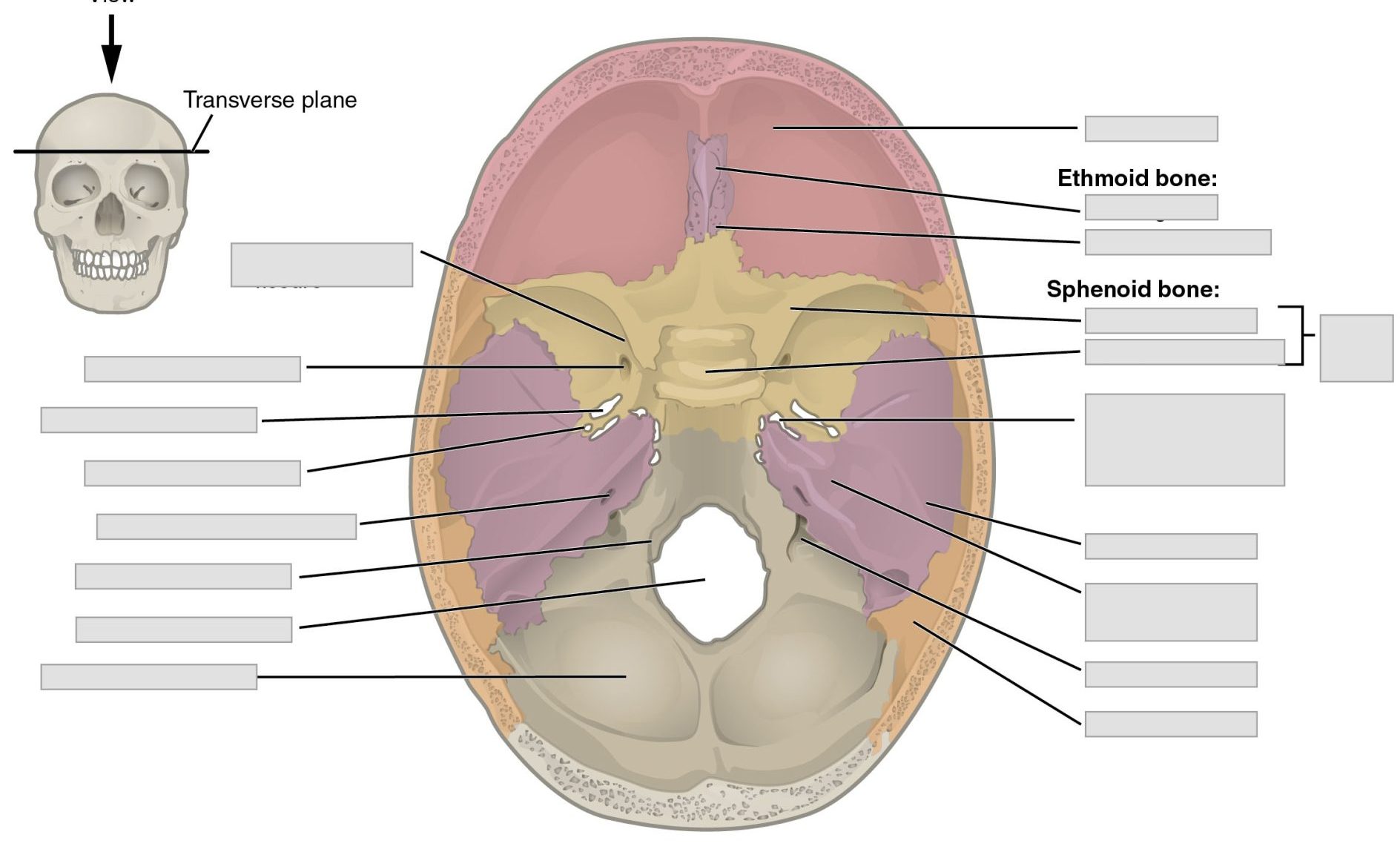
Exercise 5 Vertebral Column
Required Materials
- Vertebral column model
- Vertebra on model or articulated skeleton model
- Vertebra from the disarticulated skeleton
- Poster of the skeletal system
Procedures
- Obtain a model of vertebral column and observe its structures (Figure 7.7 and 7.8).
- Identify the structures on the list below and label them on the given vertebral column and vertebra below.
· Body · Sacral curvature · Cervical curvature · Sacrum · Cervical vertebrae · Spinous process · Coccygeal vertebrae · Superior articular process · Lamina · Thoracic curvature · Lumbar curvature · Thoracic vertebrae · Lumbar vertebrae · Transverse process · Pedicle · Vertebral foramen

Exercise 6 Thoracic Cage
Required Materials
- Thoracic cage model
- Articulated skeleton
- Poster of the skeletal system
Procedure
- Obtain a model of thoracic cage or the whole skeleton and identify its parts (Figure 7.9).
- Identify the structures on the list below and label on the figure given.
· Costal cartilage · False ribs · Floating ribs · Intercostal space · Ribs 1-12
Post-laboratory Questions
- If you are observing the skull from above a person’s head, what are the bones and structures you would be able to see? List these and describe their locations with respect to each other using anatomical terms.
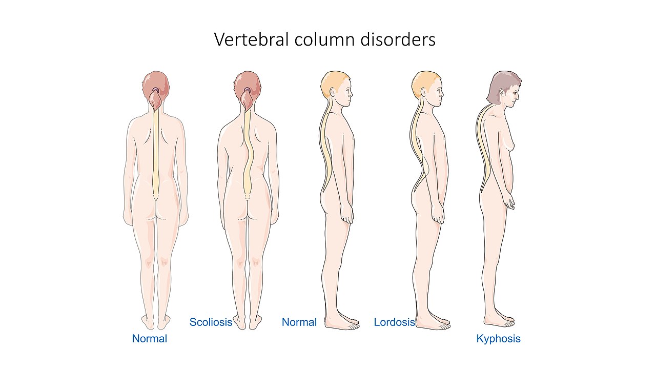
- Scoliosis, lordosis, and kyphosis are disorders of the vertebral column shown in the firs part of this chapter. Examine the figure given.
- For each disorder, identify the part of the vertebral column shown to be affected.
- How many vertebrae are in each of these regions you ID’d for each disorder?
- In open heart surgery, the sternum is cut through and ribs are spread apart to access the heart.
- List the regions of the sternum cut (from neck to belly).
- By cutting the sternum, which ribs would be affected directly? Indirectly?
