
15 Chapter 15 The Autonomic Nervous System
By Krishnan Prabhakaran
Motivation
Abnormal functioning of the autonomic nervous system can be life threatening and there is a specific term for it: dysautonomia. Dysregulation of the autonomic nervous system can produce the apparent malfunction of the organs it regulates. For this reason, dysautonomia patients often present with numerous, seemingly unrelated maladies.
Symptoms are wide ranging and can include problems with the regulation of heart rate, blood pressure, body temperature and perspiration. Other symptoms include fatigue, lightheadedness, feeling faint or passing out (syncope), weakness and cognitive impairment.
Autonomic dysfunction can occur as a secondary condition of another disease process, like diabetes, or as a primary disorder where the autonomic nervous system is the only system impacted. These conditions are often misdiagnosed.
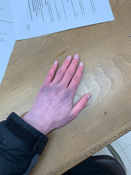
Over one million Americans are impacted with a primary autonomic system disorder. The more common forms of these conditions include Postural Orthostatic Tachycardia Syndrome POTS / Orthostatic Intolerance OI (Figure 15.1) , Neurocardiogenic Syncope NCS, Pure Autonomic Failure PAF and Multiple Systems Atrophy MSA.
Learning Objectives
Upon completion of the work in this chapter students should be able to:
- Compare and contrast the sympathetic and parasympathetic divisions of the nervous system
- Differentiate between the functions, target organs and neurotransmitters used by the sympathetic and parasympathetic divisions of the autonomic nervous system
Background.
The Autonomic Nervous System
The autonomic nervous system is tied into emotional responses and the fight-or-flight response sounds like a panic attack. In the modern world, these sorts of reactions are associated with anxiety as much as with response to a threat. It is engrained in the nervous system to respond like this. In fact, the adaptations of the autonomic nervous system probably predate the human species and are likely to be common to all mammals, and perhaps shared by many animals. However, the autonomic nervous system is not just about responding to threats. Besides the fight-or-flight response, there are the responses referred to as “rest and digest”. The digestive system has a big job to do. Much of the function of the autonomic system is based on the connections within an autonomic, or visceral, reflex.”
The Autonomic Nervous System at Work
Jayla is sitting, having an outdoor lunch with friends when a large spider lands on her plate. She immediately freezes, the food in her mouth begins to feel like a wad of dry hay, and she nearly gags as she tries to swallow it. She feels her heart race and pounding in her chest. After swallowing her food, it seems stuck in her throat and chest.
In this scenario, Jayla had been sitting, relaxed and enjoying a meal. In this relaxed state, the body would have a heart rate that is at rest, active peristalsis (activity in muscles of the digestive system), ample activity in salivary glands and in digestive gland secretions, and bronchi that are not dilated. In this relaxed state, food can be easily processed due to ample amounts of saliva and digestive enzymes in the saliva released by the salivary glands into the mouth. Salivation also facilitates swallowing by providing lubrication to the back of the throat and the esophagus. In this relaxed state, digestive fluids and enzymes in the intestines are actively produced and secreted so that food can be further processed and broken down (catabolized) for absorption of nutrients and glucose. While relaxed, Jayla’s heart beats imperceptibly and her breathing is deep.
With the sudden appearance of the spider, the rate of Jayla’s heart beat becomes more rapid, and it contracts more powerfully. In this vigilant state, Jayla senses her rapid heart rate as well as the increased force of the contraction of her heart. She also senses a shift to rapid, shallow breathing that she tries to control. Her food seems lodged near the back of her throat as she struggles to swallow her food safely.
Within seconds of seeing the spider, Jayla’s body systems shifted from reflecting calm to a state of panic and hyper-vigilance. These responses to the external threat prepare Jayla to either fight or flee the situation. In the hyper-vigilant state, Jayla’s pupils widen, she sweats and more blood is pumped through her blood vessels which permits more blood, chemicals, and hormones to flow to her skeletal muscles and respiratory system. As you can see, the effects on organ systems when in either of the states, relaxed or hyper-vigilant, are nearly opposite. These two states are controlled by two subsets of neural pathways that are part of the peripheral nervous system.
The autonomic nervous system is divided into the sympathetic nervous system and the parasympathetic nervous system. It is the complementarity of these two latter branches of the autonomic nervous system that drove the physiological changes in the “Spider Sat Down Beside Her” scenario above.
As evident from the impact of the sight of the spider on Jayla’s ability to eat her meal, activity in the parasympathetic system is associated with a relaxing meal; on the other hand, activity in the sympathetic system is associated with alertness and vigilance. In keeping with the complementary functionality of the two systems, they have been given nick names. The parasympathetic branch works for “rest and repose” (also commonly known as “rest and digest”); the sympathetic branch is known for the “fight or flight” response (variously also known as “fight, flight or freeze;” “hyperarousal;” “acute stress”).
Comparison of Somatic Motor and Autonomic Nervous Systems
| FEATURE | SNS | ANS |
| EFFECTOR | Skeletal muscle | Glands, smooth and cardiac muscle |
| GENERAL FUNCTION | Conscious or unconscious | Unconscious, affected by mental processes |
| EFFECTOR RESPONSE | Skeletal muscle contraction | Effector stimulated or inhibited |
| NEURON ARRANGEMENT (CNS TO EFFECTOR) | One only | Two: CNS to autonomic ganglion (preganglionic neuron) and ganglion to effector (postganglionic neuron) |
| CELL BODY LOCATION | Motor nuclei of cranial nerves or ventral horn of spinal cord | Preganglionic in autonomic nuclei of cranial nerves or lateral horn of spinal cord; postganglionic in autonomic ganglia |
| NUMBER OF SYNAPSES | One: Between motor neuron and effector | Two: Between pre- and postganglionic neurons and between postganglionic neuron and effector |
| AXON MYELINATION | Yes | Preganglionic = yes; Postganglionic = no |
| NEUROTRANSMITTER | Acetylcholine | Preganglionic neurons = acetylcholine
Postganglionic neurons = acetylcholine or norepinephrine |
| RECEPTOR TYPE | Nicotinic | Autonomic ganglia = nicotinic
Effectors = muscarinic for acetylcholine; α– or ß-adrenergic for norepinephrine |
General Information on the Autonomic Nervous System
- Consists of motor (=efferent) neurons and peripheral nerves that transmit to visceral effectors of the body (e.g., glands, smooth muscle, cardiac muscle)
- Involves sensory neurons that function for somatic division also
- Acts automatically and involuntarily
- Functions primarily to maintain homeostatic conditions (mainly by autonomic reflexes, although emotions can have effects also)
- Composed of organizational network of ganglia which serve as synaptic centers between preganglionic neurons with cell bodies in CNS (transmit from CNS to ganglia) and postganglionic neurons with cell bodies in ganglia (transmit from ganglia to effectors)
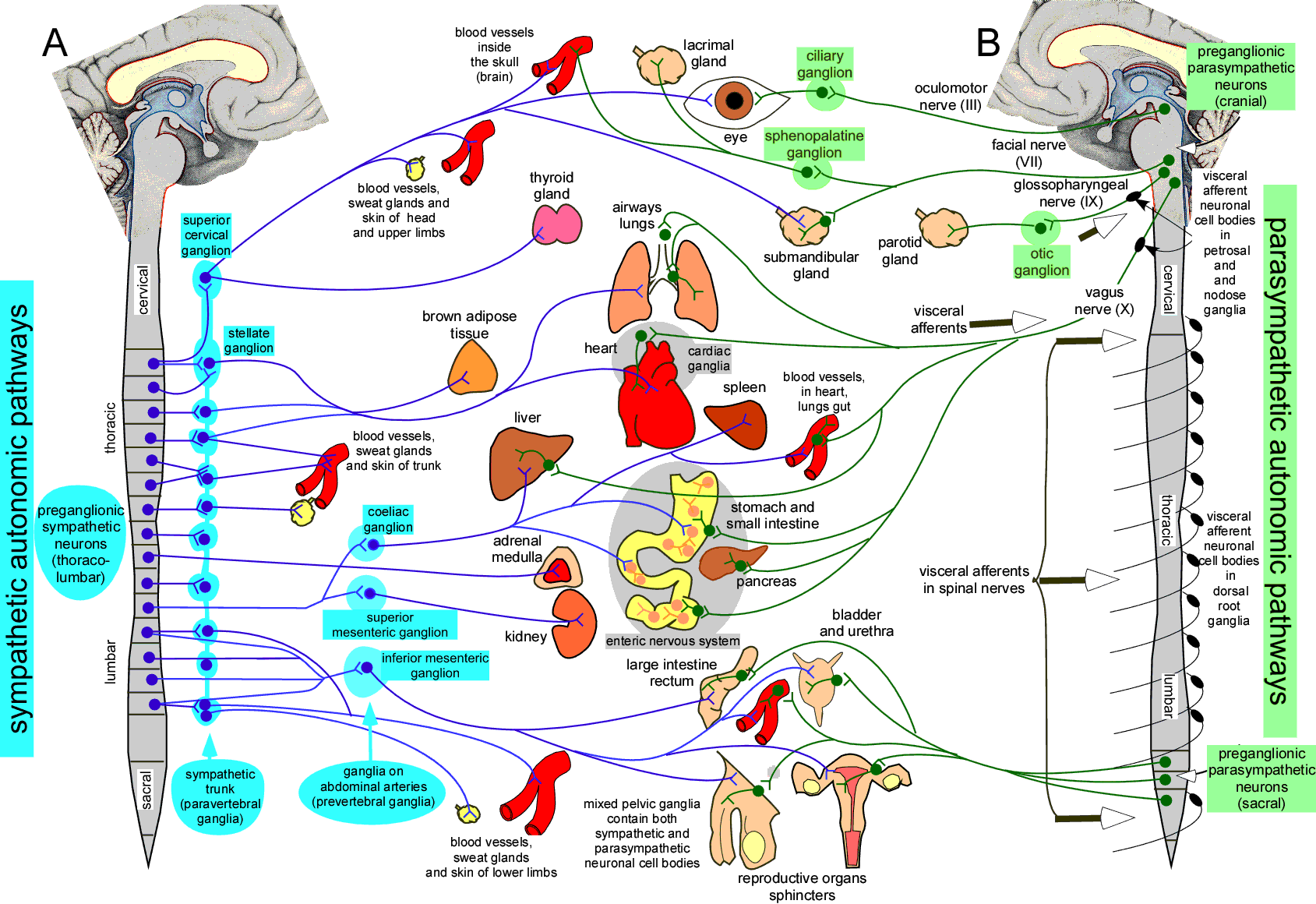
Divisions of the Autonomic Nervous System
A. Sympathetic (=thoracolumbar) Division
General information
- “Fight-or-flight” (=energy expenditure) division
- Arises from all thoracic and first two lumbar spinal nerves
General functions
- Stimulates: heart rate, intestinal sphincter constriction, urinary bladder relaxation, dilation of pupils of eyes, sweat secretion, erection of hair
- Inhibits: stomach and intestinal movements, bronchial muscles
Anatomy
- Sympathetic ganglia (=vertebral) = lie on either side lateral to ventral surface of spinal cord from second cervical vertebra to coccyx (resembling a chain of beads)
- Preganglionic neuron axons (between spinal cord and ganglion) small in diameter and myelinated = white ramus communicants
- Usually synapse with several postganglionic neurons
- Possible routes of exit of sympathetic axons
- Spinal nerves
- Preganglionic axons synapse with postganglionic neurons at same level their axons enter sympathetic chain–or–may pass superiorly or inferiorly and synapse with postganglionic neurons at different levels from where their axons enter the chain
- Postganglionic axons leave chain and pass through gray ramus communicants and reenter spinal nerve
- Innervate sweat glands, smooth muscle in blood vessels of skin or bones, and arrector pili in skin
- Sympathetic nerves
- After preganglionic neurons synapse as described for spinal nerves, postganglionic axons leave chain in a sympathetic nerve
- Innervate heart muscle, thoracic blood vessel smooth muscle, smooth muscle of esophagus and lungs and, from a sympathetic nerve plexus near the carotid artery, the head and neck sweat glands, salivary glands, smooth muscle in blood vessels, the eye, and arrector pili
- Splanchnic nerves
- Some preganglionic axons originating at T5-12 enter chain ganglia but exit at the same or different level, without synapsing, as splanchnic nerves going to collateral (=prevertebral) ganglia to synapse with postganglionic neurons
- Collateral ganglia are in the abdomen close to where major arteries (for which they are named) arise from the abdominal aorta: celiac, superior mesenteric, and inferior mesenteric
- Innervate abdominopelvic structures: smooth muscle in walls of blood vessels and organs or glands (e.g., pancreas, liver, prostate)
- Innervation of adrenal medulla
- Composed only of preganglionic neurons whose axons synapse with cells of adrenal medulla
- Eighty percent of cells secrete epinephrine and twenty percent secrete norepinephrine into blood stream
- Enhances and prolongs effect of sympathetic division; permits effect to reach organs otherwise not involved (due to lack of sympathetic innervation)
- Spinal nerves
B. Parasympathetic Division (=craniosacral)–think “vagus”
General information
- “Repose-and-repair” (=restorative)
- Arises from brainstem nuclei and S2-S4
General functions
- initiates effects generally antagonistic to sympathetic division
Anatomy
- Parasympathetic (=terminal) ganglia = lie in or near their effectors (preganglionic axons tend to synapse with only one or few postganglionic neurons)
- Nerves
- Oculomotor = smooth muscles in eyes
- Facial and glossopharyngeal = salivary glands
- Vagus (contains 75% of all parasympathetic fibers) = heart, lungs, esophagus, stomach, pancreas, liver, small intestine, and upper colon
- S2-S4 = urinary bladder, lower colon, rectum, and reproductive organs
Physiology of the Autonomic Nervous System
Neurotransmitters
- Acetylcholine
- Secreted by cholinergic axons
- Includes all autonomic preganglionic axons and all parasympathetic postganglionic axons
- Norepinephrine
- Secreted by adrenergic axons
- Includes nearly all sympathetic postganglionic axons (Exception = some that innervate sweat glands)
- *Effectors break down norepinephrine and epinephrine more slowly than acetylcholine; therefore, the effects of these neurotransmitters last longer than acetylcholine.
Receptors
-
- Cholinergic = respond to acetylcholine
- Nicotinic
- Always leads to excitatory response
- All autonomic postganglionic neurons and skeletal muscle cells
- Nicotinic
- Cholinergic = respond to acetylcholine
-
-
- Muscarinic
- may be excitatory (g., smooth muscle in wall of stomach) or inhibitory (e.g., heart muscle)
- Muscarinic
-
-
- Adrenergic = respond to norepinephrine or epinephrine
- o Types
- Alpha (α)
- Beta (ß) = more sensitive to epinephrine than to norepinephrine
- *Beta blockers bind beta receptors to reduce rate and strength of heart contraction; this, in turn, reduces blood pressure.
- o Responses = may be inhibitory or excitatory in either case
- Norepinephrine excitatory at beta receptors in heart muscle but inhibitory at beta receptors in stomach wall smooth muscle
- Epinephrine from adrenal medulla stimulates both alpha and beta receptors about equally, but norepinephrine stimulates alpha more than beta
Pre-Laboratory Questions
-
The sympathetic nervous system is referred to as the thoracolumbar system because its postganglionic neurons lie in the lateral horn of the spinal cord at thoracic and lumbar levels.
-
Preganglionic neurons of the parasympathetic nervous system.
-
Postganglionic neurons of the sympathetic nervous system receive input from all of the following sources except
-
A pre-ganglionic neuron of the sympathetic nervous system will synapse in the dorsal root ganglion chain.
-
Postganglionic neurons are arranged as a chain along side the spinal vertebral column
Exercises
- Exercise 1 Identification of the autonomic divisions
- Exercise 2 Problem solving: Acetylcholine’s autonomic effects
Exercise 1: Identification of the autonomic divisions
Place an “X” to show which division is involved for each of the following:
| CONDITION | SYMPATHETIC | PARASYMPATHETIC |
| Adrenergic fibers | ||
| Postganglionic fibers cholinergic | ||
| Long preganglionic/short postganglionic axons | ||
| Short preganglionic/long postganglionic axons | ||
| Derived from cranial and sacral nerves | ||
| Derived from spinal nerves T1 to L3 | ||
| Repose-and-repair division | ||
| Fight-or-flight division | ||
| Control more specific | ||
| Dry mouth and bronchiolar dilatation | ||
| Constricts eye pupils and decreases HR |
MATCH the following to sympathetic (S) division, parasympathetic (P) division, (B) both, or (N) neither:
- ______ Terminal ganglia
- ______ Craniosacral outflow
- ______ Two-neuron efferent chain
- ______ Adrenergic fibers
- ______ Cervical ganglia
- ______ Otic and ciliary ganglia
- ______ Increases HR, respiratory rate, and BP
- ______ Increases gastric motility and lacrimal secretion
- ______ Active when you are relaxing in a hammock
- ______ Innervation of skeletal muscles
- ______ Active when you are competing in the Boston Marathon
- ______ Nerve cell bodies in ganglia
Exercise 2: Problem solving: Acetylcholine’s autonomic effects
We have a problem solving activity using a patient’s case and this is presented in multiple-choice format.
Consider each choice individually and write an argument for accepting or rejecting it. Since the problem has one best answer, you will write one argument for acceptance and four for rejection.
For each response, first state whether you are accepting or rejecting that statement. Then, write a detailed explanation of why you accept or reject each of the choices.
PROBLEM:
Mr. A. Prentice has been suffering from functional urinary retention and a hypoactive urinary bladder. Bethanechol, a drug that mimics acetylcholine’s autonomic effects, is prescribed to manage his problem. Which of the following adverse effects might Mr. A. Prentice experience while taking this drug?
A. Dry eyes due to deficient tear formation.
B. Deficient salivation.
C. Constipation.
D. Decreased sexual arousal.
E. Diarrhea
Post-laboratory Questions
-
A presynaptic parasympathetic neuron may reach its target organ by which mechanism?
-
Sympathetic signals to smooth muscles of blood vessels of arms and legs will be sent via which neurotransmitters?
- The effects of the sympathetic nervous system are often more generalized and widespread than the effects of the parasympathetic nervous system (which tend to be more specific and localized). How does the anatomy of the sympathetic nervous system provide for more widespread effects?
4. Why do we feel cold when facing a “fight or flight” situation?
5. Describe major parasympathetic and sympathetic physiological effects on target organs.
Abnormally fast heart rate
