
23 Chapter 23 The Digestive System
Motivation.

Diabetes is a disorder of pancreas which is an accessory organ of the digestive system. In diabetes the pancreas is unable to regulate the level of sugar in our blood. Diabetes prevalence is disparately high in African-American populations in the U.S. compared to other racial groups.
The pancreas is an associated part of the digestive system that consists of the mouth, pharynx, esophagus, stomach, small and large intestine. Other associated components of the digestive system are the liver and gall bladder. The digestive system functions to process food that is eaten and to convert it into useable energy by metabolism and therefore the digestive system is important in our lives for survival. Diabetes is a disorder that can be managed with a proper diet and exercise. In this chapter, you will learn the parts of the digestive system and its associated organs and their functions.
Learning Objectives
Upon completion of the work in this chapter students should be able to:
- Identify the structures of the digestive system and associated parts.
- Describe the histology of the esophagus, stomach, small and large intestine.
- Explain the function of the stomach, small and large intestine.
- Describe the function of the liver, gall bladder and pancreas.
- Explain carbohydrate metabolism.
Background.
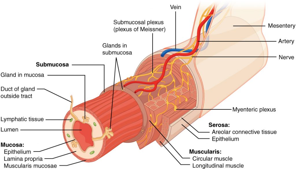
The digestive system consists of the mouth, pharynx, esophagus, stomach, small intestine and large intestine. The organs associated with the digestive system are the liver, gall bladder and pancreas and several glands (Figure 23.2). Each organ of the digestive system is made up of specialized tissues with specific functions (Figure 23.3).
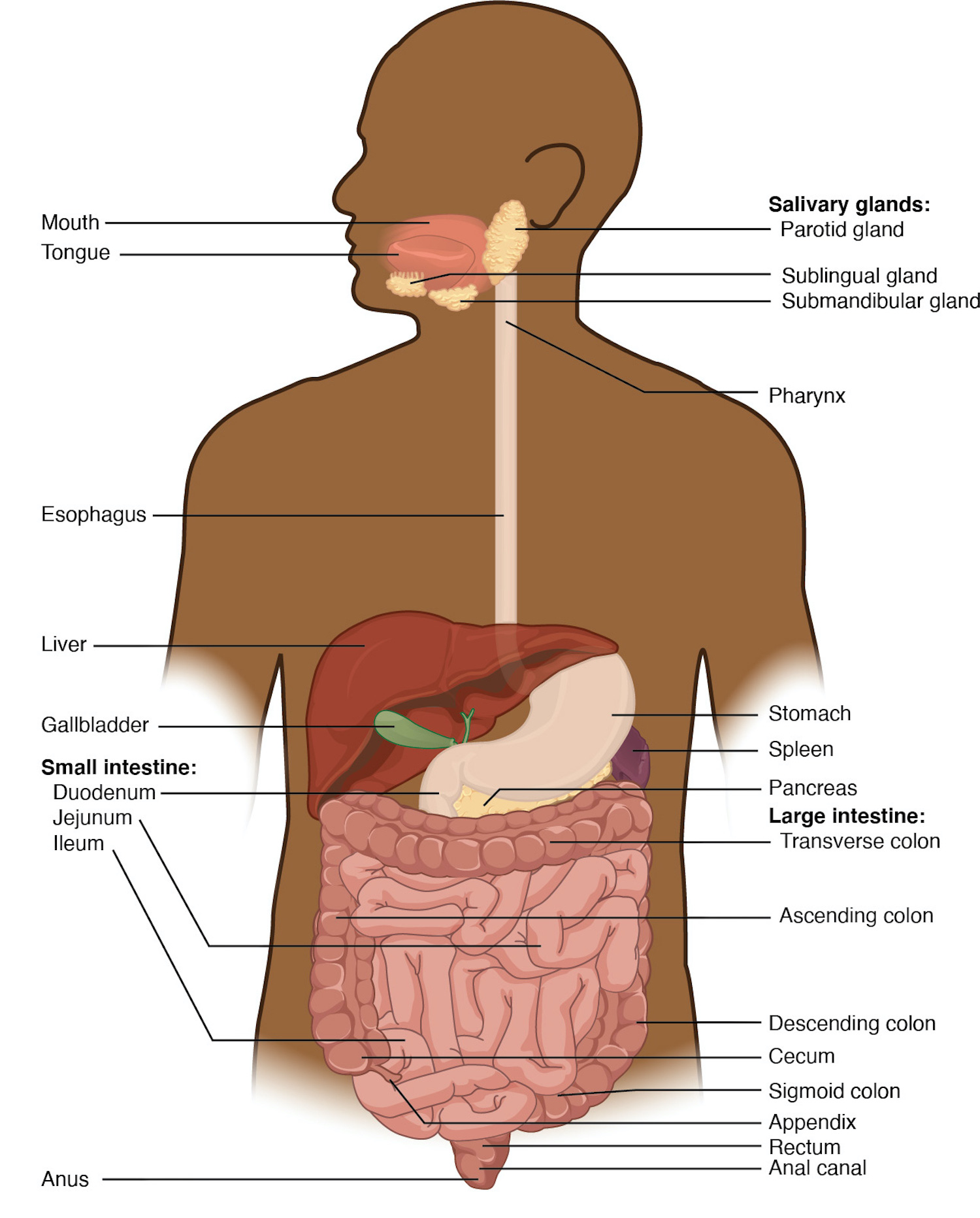
The digestive system is mostly suspended in the abdominal cavity. Food goes through the digestive canal or alimentary canal (Figure 23.2) and is processed as it moves down the lumens of a set of digestive organs starting with the mouth.
The mouth is a cavity with teeth, tongue and salivary glands. The pharynx is a hollow tube that connects with the esophagus (Figure 23.4). (The pharynx is also connected with the respiratory system.)
The tubular esophagus passes through an opening in the diaphragm (muscular organ that divides the thoracic and abdominal cavity.) It leads to a curved stomach which is essentially a holding bag that is a muscular organ (Figures 23.4 and 23.5).
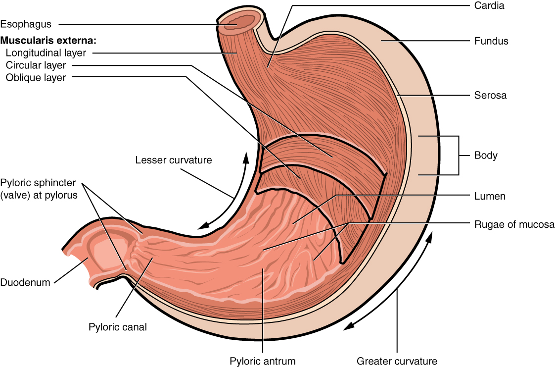
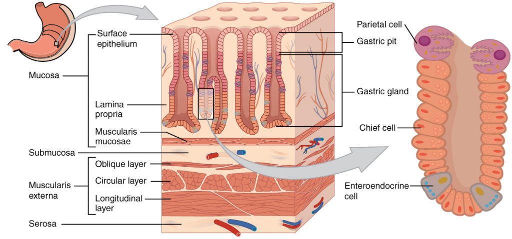
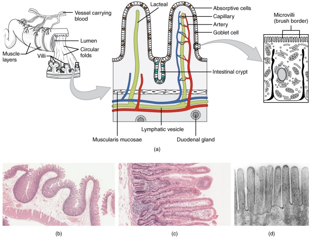
The stomach is connected to the small intestine which is approximately 671 cm., long. Most of digestion and absorption occur in the small intestine. Between the stomach and the small intestine is a pyloric sphincter. It is a valve that prevents the backflow of food once it passes from the stomach into the small intestine (Figure 23.6).
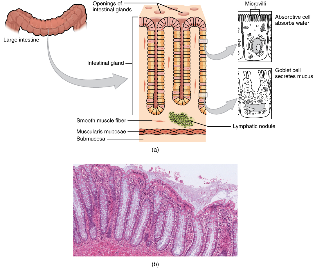
Undigested food passes from the small intestine into the large intestine. The large intestine is 213 cm long (Figure 23.7). The large intestine stores waste such as feces and water.
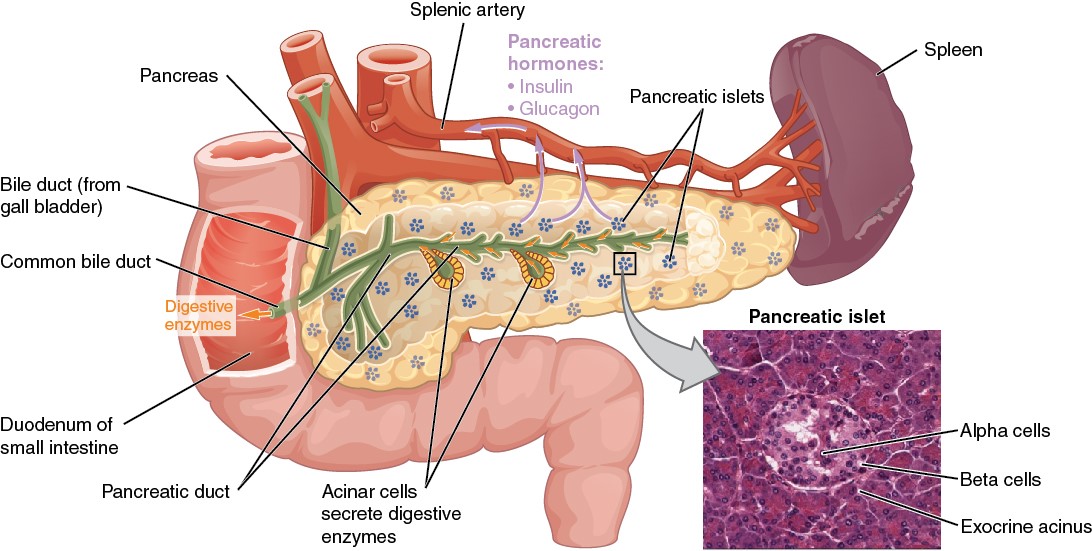
The pancreas is an organ located in the bend between the stomach and small intestine (Figure 23.8). It secretes a number of enzymes that mediates to break down carbohydrates, proteins and fats.
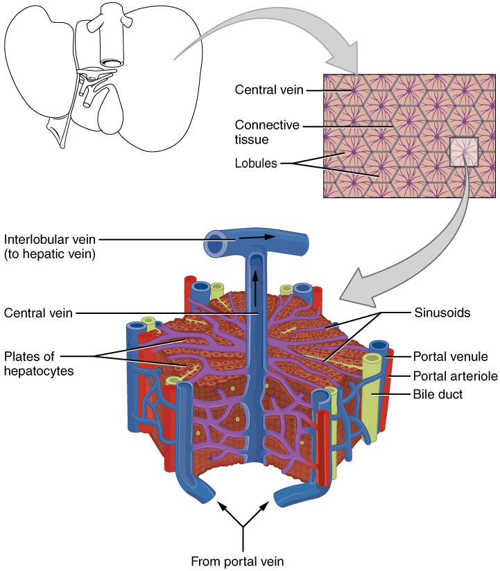
The liver functions to remove toxins and store sugar, as well as producing bile that is stored in the gallbladder and helps with digestion of fats (Figure 23.9). The gall bladder stores this bile that emulsifies fats.
The main functions of the digestive system are movement food (propulsion), digestion of food by secreted chemicals or mechanically, and absorption of the digested food (Figure 23.10, Table 1).
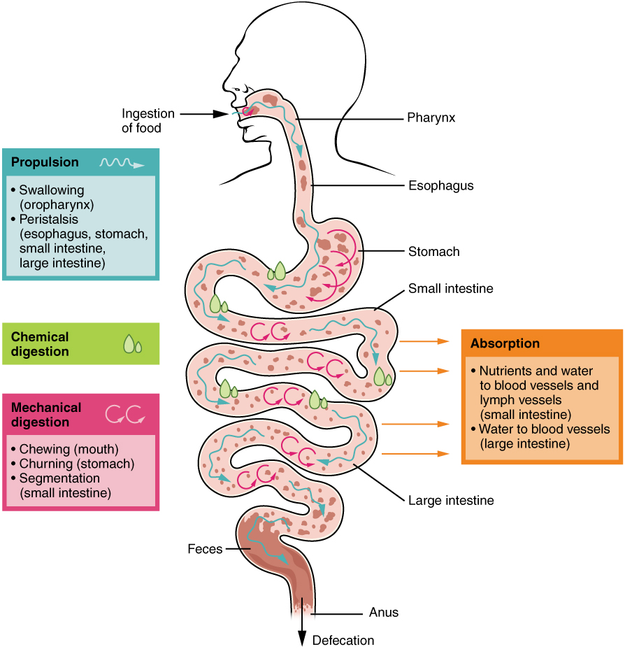
Table 1 Functions of the Digestive System
| Organ | Major functions | Other functions |
| Mouth |
|
|
| Pharynx |
|
|
| Esophagus |
|
|
| Stomach |
|
|
| Small intestine |
|
|
| Accessory organs |
|
|
| Large intestine |
|
|
Pre-Laboratory Questions
- Relate the parts of the digestive to your own body. Name the structures in the mouth that are associated with the digestive system.
- Draw and name the parts of the stomach.
- The stomach functions to ___________.
- Look up the histology of the stomach, small and large intestines. What four structures do these organs have in common?
- In the abdominal cavity, the liver is located in the ______ ______ quadrant. Name two functions of the liver.
Exercises
- Exercise 1 Main organs of the digestive system
- Exercise 2 Accessory organs of the digestive system
- Exercise 3 Histology of esophagus, stomach, small intestine and large intestine (optional)
- Exercise 4 Microanatomy of the pancreas and liver (optional)
- Exercise 5 Digestion of starch by salivary enzymes
Exercise 1 Main organs of the digestive system
Required Materials
- The Digestive System Poster
- Human Digestive System Model
- Torso Models
- Post-it notes
- Labeling tape
Procedure
- Study a poster and models of the digestive system and identify the mouth, pharynx, esophagus, stomach, small and large intestine.
- Using post-it notes or labeling tape label these structures on the torso model. Take a picture and insert the picture in the space below. Alternatively you can sketch and label these structures.

Exercise 2 Accessory organs of the digestive system
Required Materials
- The Digestive System Poster
- Human Digestive System Model
- Torso Models
- Construction paper
- Tape
- Markers
Procedure
- On your torso model, locate the precise position of the organs associated with the digestive system: pancreas, liver, gall bladder.
- The abdominal cavity can be divided into four quadrants or nine regions. Use these quadrants/ regions to locate the organs. Use colorful construction paper, tape and markers to delineate the four quadrants of the abdominal cavity on the model.
- Write in the pancreas, liver, and gallbladder into the quadrants you find them in. Take a picture of your labeled model. Insert the picture below. Alternatively, you can sketch and label to show the abdominal locations of these accessory organs.
- Fill in the chart below indicating the location of the digestive organs and the accessory organs of the digestive system that reside in the abdominal cavity: stomach, small intestine, large intestine, liver, gall bladder, pancreas.
| Right Upper Quadrant (RUQ)
|
Left Upper Quadrant (RUQ)
|
| Right Lower Quadrant (LLQ)
|
Left Lower Quadrant (LLQ)
|
Exercise 3 Histology of esophagus, stomach, small intestine and large intestine (optional)
Required Materials
- Compound microscope
- Microscope lens paper
- Microscope lens cleaning solution
- Microscope immersion oil
- Slide of Mammal Esophagus
- Mammal Stomach Composite slide
- Mammal Intestine Composite slide
- Large Intestine (Human) slide
Procedure
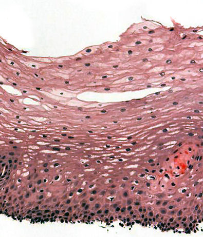
- Obtain histological slides and study the structure of the esophagus, stomach, small and large intestine.
- There are four structures that you should be able to identify by looking through a microscope: mucosa, submucosa, muscularis and serosa.
- Study the tissues and record how they are different. Provide labeled sketches below to show these layers in the four slides:
Exercise 4 Microanatomy of the pancreas and liver (optional)
Required Materials
- Compound microscope
- Microscope lens paper
- Microscope lens cleaning solution
- Microscope immersion oil
- Human Pancreas slide
- Human Liver slide
Procedure
- Obtain slides to show the tissues and cells that make up the pancreas and liver. These are accessory organs of the digestive system.
- Locate the islets of Langerhans that play a critical role in controlling blood sugar levels. What is the hormone that is secreted by the islets of Langerhans? ______________
- The liver is divided into lobules. What are the cells that make up the liver primarily? State their function. _________________________________
- Sketch what you observed under the microscope in the pancreas and liver slides. Label the hepatocytes of liver, and the islets and exocrine acini of pancreas.
Exercise 5 Digestion of starch by salivary enzymes
Required Materials
- Test tubes
- Test tube rack
- Tube labeling pen
- 1% starch solution
- Water
- Iodine (KI) solution in dropper bottle
- Graduated cylinder
- ruler
Procedure
- Carry out an experiment to show the presence of carbohydrates. Ask your instructor for three test tubes.
- Label test tubes A, B & C. The test tubes A & B will be your experimental sample. Test tube C will be control.
- Test Tube A: Saliva + Starch
- Test Tube B: Water + Starch
- Test Tube C: Water + Water
- Collect saliva in test Test Tube A.
- Use a ruler to measure the amount of saliva in Test Tube A.
- Mark Test Tube B and C to the same level. Add water to this level.
- Put 2 ml of starch into Test Tube A and into Test Tube B.
- Put 2 ml of deionized water into Test Tube C.
- Put a drop of iodine (KI) solution into each tube. Remember that iodine solution itself is yellow/brown and it turns purple in the presence of starch.
- Write down the results in the table below. Put a check mark if you see a reaction (purple) and an X if there is none.
| Test Tube A | Test Tube B | Test Tube C | |
| Reaction |
10. Did your saliva digest starch? Use your results to explain your answer. ____________________________
Post-laboratory Questions
- Name the sphincter muscles that allow foods to enter and leave the stomach.
- One role of hydrochloric acid HCl in the stomach is to convert a precursor enzyme ________ to ________.
- The four layers of the small and large intestine are _________, _______, _______, and _______.
- The islets of Langerhans secretes a hormone called_________ that regulates blood sugar levels.
