
10 Chapter 10 Muscle Tissue
By Joseph D’Silva
Motivation.
Muscles really stand out in athletes. You cannot miss them. The athletes we see here are Shaq O’Neil, Serena Williams and LeBron James. Their skeletal muscles stand out as you can see from the pictures below.

But athletes suffer injuries to their muscles. You can relate to them because you have seen them carried off the court or field on stretchers. Think of an athlete you have seen because he injured a hamstring muscle! Sports medicine and physiotherapy are huge nowadays. As more and more athletes compete, there will be more muscle injuries and you can contemplate building a career in sports medicine for yourself.
Learning Objectives
Upon completion of the work in this chapter students should be able to:
- Identify and define muscle fiber.
- State the organelles located in the muscle fiber.
- Compare and contrast skeletal, cardiac and smooth muscle fibers.
- Define neuromuscular junction and state its function.
Background.
We have skeletal muscles in our own bodies (maybe not as big as the athletes’ above!). The flesh we are made up of contains muscle tissue and is attached to bones. Muscles are needed to move our body parts. They move our skeleton, heart and gastrointestinal tract. There are three types of muscles: skeletal, cardiac and smooth. Cardiac muscle is in our heart and smooth muscles are located in the walls of our stomach and intestine as well as other tubular organs.
Skeletal muscle functions to help us to move bones. We can will them to do that voluntarily. However, cardiac and smooth muscles are beyond our control and function involuntarily from the time we are in the womb until we pass away. Skeletal muscles need our attention for many reasons. One of them is to maintain posture. Also, to lift weights, anchor ourselves, plant our feet. With age, muscle tone can be lost. Particularly, the elderly skeletal muscles need attention because they do not use their muscles as much as they should resulting in muscle loss. There are also disorders of the musculo-skeletal system that can be debilitating.
To know about muscles, you have to study their histology first. That is, you have to look at muscle tissue under the microscope. Once you know that, you can study the large muscles that stand out in our body. In this chapter, you will learn to identify skeletal, cardiac and smooth muscles.
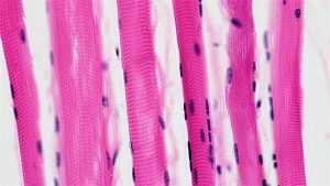
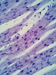
Skeletal muscles contain tissues that are made up of muscle cells aka muscle fiber (Figure 10.2).
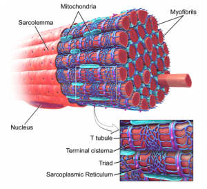
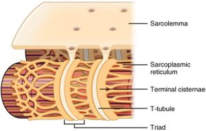
Each muscle fiber has a cell membrane (sarcolemma), cell cytoplasm (sarcoplasm) and nucleus. The cytoplasm is dotted with organelles (Figure 10.3): Golgi apparatus, endoplasmic (sarcoplasmic) reticulum, mitochondria, ribosomes, lysosomes, myofibrils and myofilaments, triads, A-band, I-band (Figure 10.4 and 10.5). Each of the many organelles has a particular function. But as a whole they produce relaxation and contraction in muscle tissue.
The sarcolemma is the membrane that covers the muscle cell (Figure 10.3). The cell contains cytoplasm in which myofibrils are very prominent. The myofibrils are surrounded by mitochondria and endoplasmic reticulum. There are three structures around the myofibrils that form the triad: terminal cisterna, T-tubule and sarcoplasmic reticulum (Figure 10.4). The myofibrils contain two protein structures known as myosin and actin. The sarcomere is a unit of structure and function in the muscle cell. It runs from one I-band to another with the A-band in the middle. The I-band and A-band can only be observed with an electron microscope. The A-band is dark. The I-band is light (Figure 10.5). When the sarcomere shortens reducing the distance between the two Z discs, the muscle contracts (Figure 10.5).
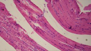
There are structural and functional differences between the three types of muscles. Skeletal muscle fibers can be identified by their multinucleate cytoplasm and striations (Figure 10.2) . Skeletal muscles move bones. Cardiac muscle fibers have one nucleus per cell, branched striations and intercalated disk (Figure 10.6). Cardiac muscles are found in the heart where they squeeze it to pump blood. Smooth muscles do not have striations. They are spindle-shaped with one nucleus in each cell (Figure 10.7). Smooth cells are found mostly in the gastrointestinal tract where they move the stomach and intestine as well as in the walls of other tubular organs such as the urinary tract and reproductive tract, helping move materials along within these tubes.
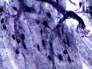
The nervous system and muscular system work closely together to maintain proper muscle contraction and relaxation. Skeletal muscles are innervated by motor neurons we can voluntarily activate or inactivate (Figure 10.8). The axon ends from the neuron send signals to the motor end plate of the sarcolemma of the skeletal muscle fiber to contract and relax the muscles. A neurotransmitter (chemical) called acetylcholine is released from the axon into the space between the neuron and muscle fiber (synaptic cleft). The neurotransmitter then binds cell surface proteins on the sarcolemma to initiate events for muscle contraction.
Pre-Laboratory Questions
- The sarcomere is a unit of structure in the muscle fiber. Describe the structure of a sarcomere. Use diagrams and electron micrographs to help you.
- The sarcomere is also a unit of function. How can you relate the function of the sarcomere to the movement of a muscle, say biceps?
- Define a (a) skeletal muscle, (b) smooth, and (c) cardiac muscle.
- Create a concept map listing events that occur in a neuromuscular junction when dendrites receive a stimulus such as a pin prick.
Exercises
- Exercise 1. Identify the microscopic features of skeletal muscle
- Exercise 2. Identify the organelles found in a muscle fiber
- Exercise 3. Identify cardiac muscle tissue
- Exercise 4. Identify smooth muscle tissue
- Exercise 5. Describe neuromuscular junction
Exercise 1. Identify the microscopic features of skeletal muscle
Required Materials
- Compound microscope
- Skeletal muscle slide
Procedure
- Obtain a slide of the skeletal muscle (Figure 10.2)
- Using a compound microscope focus on the tissue using the 4x objective.
- Switch to the low magnification 10x objective lens and scan the slide showing skeletal muscle.
- Repeat your observations high magnification of the 40x objective.
- Notice the dark nuclei. They are more than one in each cell and are located to a side in the sarcoplasm.
- Observe the striations that appear as light and dark bands. Striations and multinuclei define skeletal muscle fiber.
- Sketch what you observed at low and high magnification and label the main structures.
Exercise 2. Identify the organelles found in a muscle fiber
Required Materials
- 3D Model of the Sarcomere
- Post-it notes
Procedure
- Observe a 3-dimensional model of the sarcomere.
- Use your post-it notes to label on the model the organelles in the sarcoplasm: sarcolemma, sarcoplasm, nucleus, sarcomere, myofibrils, myofilaments and triad (Figure 10.3, 10.4, 10.5)
- Take a picture of the model with your post-it notes attached. Insert in the space below. (Alternatively, you can draw a picture of the sarcomere and label this figure with the structures indicated.)

Exercise 3. Identify cardiac muscle tissue
Required Materials
- Compound microscope
- Cardiac muscle slide
Procedure
- Obtain a slide of the cardiac muscle (Figure 10.6)
- Using a compound microscope focus on the tissue using the 4x objective.
- Switch to the low magnification 10x objective lens and scan the slide showing cardiac muscle.
- Repeat your observations high magnification of the 40x objective.
- Notice striations, intercalated disks (bands), single nucleus in a cell. The cells are also branched.
- Sketch what you observed at low and high magnification and label the main structures.
Exercise 4. Identify smooth muscle tissue
Required Materials
- Compound microscope
- Smooth muscle slide
Procedure
- Obtain a slide of the smooth muscle (Figure 10.7)
- Using a compound microscope focus on the tissue using the 4x objective.
- Switch to the low magnification 10x objective lens and scan the slide showing smooth muscle.
- Repeat your observations high magnification of the 40x objective.
- Unlike skeletal and cardiac muscle fibers, there are no striations. There is one nucleus per nucleus.
- Sketch what you observed at low and high magnification and label the main structures.
Exercise 5. Describe neuromuscular junction.
Required Materials
- Compound microscope
- Neuromuscular spindle slide
Procedure
- Obtain a slide that shows skeletal muscle tissue with a nerve cell. The axons of nerve cells (neurons) touch the sarcolemma and stimulate it to relax and contract (Figure 10.8)
- Using a compound microscope focus on the tissue using the 4x objective.
- Switch to the low magnification 10x objective lens and scan the slide showing axon ends and motor end plate.
- Repeat your observations high magnification of the 40x objective.
- Notice the nerves, axon ends, striated muscle fibers, and the motor end plate.
- Sketch what you observed at low and high magnification and label the main structures.
Post-laboratory Questions
- Construct a table with three raws. Now, describe similarities and dissimilarities in structure (a) between skeletal muscle and smooth muscle; (b) smooth and cardiac muscle; (c) skeletal muscle and cardiac muscle.
Similarities Dissimilarities Question (a) skeletal vs. smooth
Question (b) smooth vs. cardiac
Question (c) skeletal vs. cardiac
2. Draw a muscle fiber and label:
a. Myofilament
b. Sarcoplasmic reticulum
c. T-tubule
d. Triad

3. Fill in the blanks:
a. Myofilament plays a role in _______________________
b. The sarcoplasmic reticulum functions to _____________________
c. The T-tubule can be defined as _______________________
d. The triad is a set of structures that ______________________________
4. The synaptic cleft is a _______________ between the _________________ and _______________
5. Acetylcholine is a neurotransmitter located in ____________________
6. Explain what happens when acetylcholine is released into the synaptic cleft.
7. What is the function of dendrites? What is the function of axons?
