
5 Chapter 5 The Integumentary System
By Ganesan L. Kamatchi
Motivation

Human skin is the first anatomical site a nurse examines when seeing a patient, using it as an indicator of general health such as oxygenation, nutritional status, and injury1. Skin color is mostly based on the pigmentation or melanin found in the keratinocytes in the epidermis of skin. In addition, the blood supply to the surface (redness), injury (bruising), and jaundice (yellowing) can affect skin color.
Skin color of different races has led to many misconceptions in medicine. 40% of white medical students surveyed in 2016 thought that black skin is thicker than white skin therefore making black patients more resistant to pain!2 In reality, the sensory receptors called free nerves endings found within the dermis of skin detect pain and are completely unrelated to skin color which is not just black and white, but many shades in between. Let’s get our facts right and serve patients of all skin color without misconceptions.
Credit:
- Everett JS, Budescu M, Sommers MS. Making Sense of Skin Color in Clinical Care. Clin Nurs Res. 2012;21(4):495-516. doi:10.1177/1054773812446510
- How we fail black patients in pain | AAMC. https://www.aamc.org/news-insights/how-we-fail-black-patients-pain. Accessed July 22, 2020.
Learning Objectives
Upon completion of the work in this chapter on the integumentary system, students should be able to:
- Describe the layers of epidermis of thick and thin skin
- Identify the basis for skin color
- Differentiate the layers of dermis
- Explain epidermal ridges, dermal papillae and fingerprints
- Differentiate sebaceous glands, merocrine (eccrine) and apocrine sweat glands
- Identify parts of hair follicle
- Describe the structure of nail
Background.
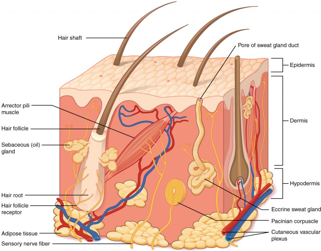
Although you may not typically think of the skin as an organ, it is in fact made of tissues that work together as a single structure to perform unique and critical functions. The skin and its accessory structures make up the integumentary system, which provides the body with overall protection. The skin is made of multiple layers of cells and tissues, which are held to underlying structures by connective tissue. The top layer is called epidermis and comprises several layers of cells. The layer underneath the epidermis is called dermis and is made of connective tissue. The dermis is well vascularized (has numerous blood vessels) and has numerous sensory, and autonomic and sympathetic nerve fibers ensuring communication to and from the brain. The layer below the dermis is called hypodermis, not considered as a part of the integument, and consists of well-vascularized, loose, areolar connective tissue and adipose tissue (Figure 5.2).
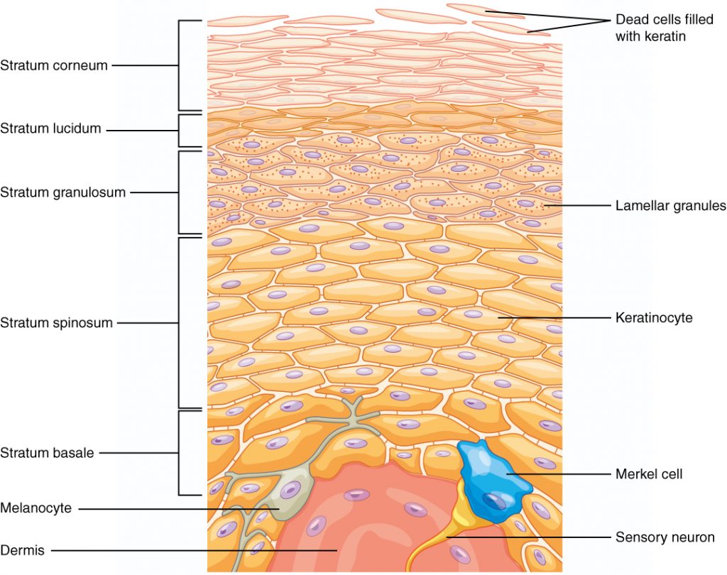
The epidermis is composed of keratinized, stratified squamous epithelium. It is made of four or five layers of epithelial cells, depending on its location in the body. Skin that has four layers of cells is referred to as “thin skin” (all over the body except the palms of the hand and the soles of the foot). From deep to superficial, these layers are the stratum basale, stratum spinosum, stratum granulosum, and stratum corneum. The palm and sole have an additional layer, stratum lucidum (between stratum corneum stratum granulosum) and referred to as the “thick skin” (Figure 5.3).
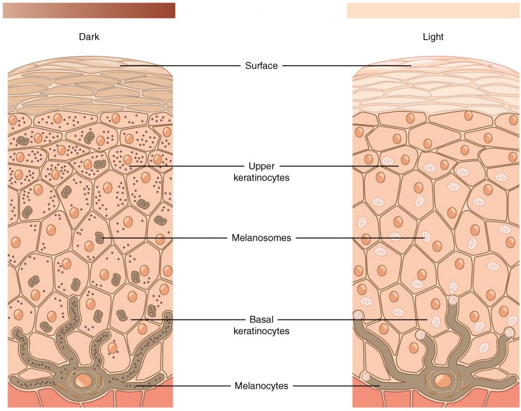
The color of skin is influenced by the presence of a pigment called melanin. It is produced by cells called melanocytes, which are found scattered throughout the stratum basale of the epidermis. The melanin is transferred into the keratinocytes via a cellular vesicle called a melanosome (Figure 5.4).
Hair
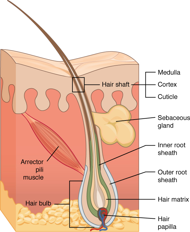
Accessory structures of the skin include hair, nails, sweat glands, and sebaceous glands. These structures embryologically originate from the epidermis and can extend down through the dermis into the hypodermis (Figure 5.5 and Figure 5.6).
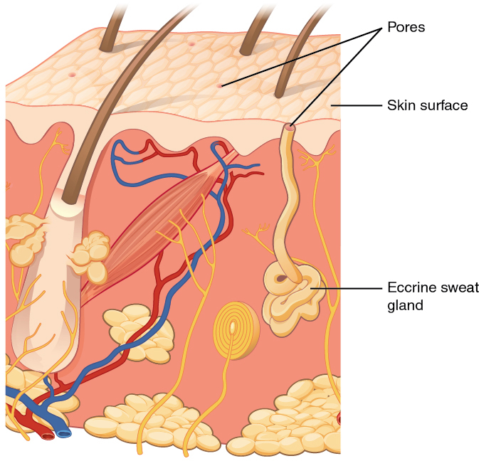
Sweat Glands
When the body becomes warm, sudoriferous glands (aka. sweat glands) produce sweat to cool the body. Sweat glands develop from epidermal projections into the dermis and are classified as eccrine sweat glands and apocrine sweat glands. The location of these glands and the type of sweat released by them are different. Eccrine sweat glands are the major sweat glands of the human body, responsible for temperature regulation and found in virtually all skin, with the highest density in palm and soles, then on the head, but much less on the torso and the extremities.
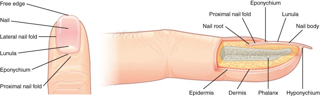
Nail
Nail is a specialized skin appendage that is a claw-like keratinous plate at the tip of the fingers and toes in most primates. Fingernails and toenails are made of a tough protective protein called alpha-keratin which is a polymer and found in the hooves, hair, claws and horns of vertebrates. The nail consists of the nail plate, the nail matrix and the nail bed below it, and the grooves surrounding it.
Pre-Laboratory Questions
Review the background information provided in the chapter and answer these questions prior to starting the exercises.
- What are the layers of skin?
- What is the basis for skin color?
- Does the skin have any sense organs?
- What are the functions of skin?
- What are the accessory structures of skin?
Exercises
- Exercise 1 Layers of Integument
- Exercise 2 Layers of Epidermis
- Exercise 3 Skin Pigmentation
- Exercise 4 Axillaries of Skin – Hair
- Exercise 5 Axillaries of Skin – Eccrine Sweat Glands
Exercise 1 Layers of Integument
Required Materials
- Slide of skin
- Classroom model of skin
Procedure
- Obtain a slide of skin or a model of skin.
- Observe the organization of various layers of the skin
- Identify
- Epidermis
- Dermis
- Hypodermis and
- Accessory structures
- Sketch the skin and label the parts of the integument shown in Figure 5.2 above, observed at low and high magnification.
Exercise 2 Layers of Epidermis
Required Materials
- Compound microscope
- Slide of thick skin (palmar or plantar skin)
- Skin slide (hairy skin, skin with sweatglands, etc)
Procedure
- Obtain a slide of either “thick” or “thin” skin.
- Place it on the stage of the microscope and scan the slide at low power.
- Once the epithelial layer is visible in the field of view, switch to high power.
- Examine all the layers of the epidermis and study the difference between the layers (Figure 5.3).
- Sketch the layers as seen in the microscope and make sure to label all structures identified, observed at low and high magnification.
Exercise 3 Skin Pigmentation
Required Materials
- Compound microscope
- Slide of pigmented skin epithelium
Procedure
- Obtain a slide of pigmented skin.
- Place it on the stage of the microscope and scan the slide at low power.
- Once the layers are visible in the field of view, switch to high power.
- Examine all the layers of the epidermis and study the pigmented layers (Figure 5.4).
- Sketch the layers as seen in the microscope and label all relevant structures, observed at low and high magnification.
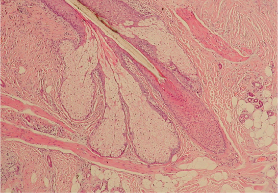
Exercise 4 Axillaries of Skin – Hair
Required Materials
- Compound microscope
- Slide of scalp skin (hairy)
Procedure
- Obtain a slide of scalp, place it on the stage of the microscope and scan the slide until a hair follicle is visible in the field of view.
- Observe that there are three distinct regions to a hair: 1) the shaft, the portion of hair that is outside the body surface; 2) the root, the portion within the skin and 3) the bulb, the enlarged base of the hair (Figure 5.5 and 5.6).
- Note the other accessory structures of skin such as the oil secreting sebaceous glands and the apocrine sweat glands that are connected to the hair root; and arrector pili muscle that is also attached to the hair bulb (Figure 5.5 and 5.6.
- Sketch the skin as seen in the microscope and label the structures related to hair as well as adjacent parts, observed at low and high magnification.
Exercise 5 Axillaries of Skin – Eccrine Sweat Glands
Required Materials
- Compound microscope
- Slide of skin from general body surface showing sweat glands
Procedure
- Obtain a slide of skin, place it on the stage of the microscope and scan the slide until an eccrine sweat gland is visible in the field of view.
- Observe that it originates from the dermis and the duct reaches the skin surface and the pore is exposed to the skin surface (Figure 5.7).
- Sketch the skin area with eccrine sweat glands as seen in the microscope, observed at low and high magnification.
Exercise 6 Axillaries of Skin – Nail
Required Materials
- Compound microscope
- Slide of developing nail
Procedure
- Obtain a slide of nail, place it on the stage of the microscope and scan it to view all the parts of the nail mentioned above
- Identify
- Nail plate
- Nail matrix
- Nail body
- Nail root
- Lunula
- Sketch the nail and its parts as seen in the microscope, observed at low and high magnification.
Post-laboratory Questions
1.The papillary layer of the dermis is most closely associated with which layer of the epidermis?
A. stratum spinosum
B. stratum corneum
C. stratum granulosum
D. stratum basal
2.Eccrine sweat glands ________.
A. are present on hair
B. are present in the skin throughout the body and produce watery sweat
C. produce sebum
D. act as a moisturizer
3.Sebaceous glands ________.
A. are a type of sweat gland
B. are associated with hair follicles
C. may function in response to touch
D. release a watery solution of salt and metabolic waste
4.Similar to the hair, nails grow continuously throughout our lives. Which of the following is furthest from the nail growth center?
A. nail bed
B. hyponychium
C. nail root
D. eponychium
5.An individual using a sharp knife notices a small amount of blood where he just cut himself. Which of the following layers of skin did he have to cut into in order to bleed?
A. stratum corneum
B. stratum basale
C. papillary dermis
D. stratum granulosum
6. As you are walking down the beach, you see a dead, dry, shriveled-up fish. Which layer of your epidermis keeps you from drying out?
A. stratum corneum
B. stratum basale
C. stratum spinosum
D. stratum granulosum
7.An individual has spent too much time sun bathing. Not only is his skin painful to touch, but small blisters have appeared in the affected area. This indicates that he has damaged which layers of his skin?
A. epidermis only
B. hypodermis only
C. epidermis and hypodermis
D. epidermis and dermis
