
11 Chapter 11 The Muscular System
By Joseph D’Silva
Motivation.
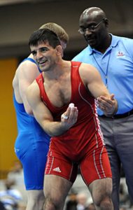
Have you ever noticed how skeletal muscles stand out in most men compared to most women? Men are more heavily built and it has to do with genetics and DNA! The build comes from those muscles that are attached to bone or bones. Skeletal muscles make up the muscular system. Each muscle has a name. For every muscle, there is a point of origin in a bone and an insertion in another. Skeletal muscles are bound to bone by tendons. But ligaments bind muscles to muscles. This is important to know. Basically, skeletal muscles move our body. But injuries such as sprain, tear, contusion can occur. A sprain is a damage to muscle fibers. In sports medicine, the patients are athletes that can suffer from tendinitis and pulled hamstrings.
Learning Objectives
Upon completion of the work in this chapter students should be able to:
- Identify facial muscles, name them and state their functions.
- Name and identify thoracic muscles.
- Identify muscles of the abdominal wall.
- Identify appendicular muscles of the pectoral girdle.
- List muscles of the lower limb.
Background.
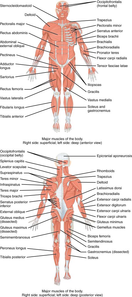
Muscles are tissues that stand out in our bodies. We are all familiar with them. You already know the name of the muscle on your forearm. It is the biceps. Two particular features of your muscles is that each one of them has a name and a function. The names identify them. Muscles bind bone to bone. Relax a muscle and the bone will extend out. Contract a muscle and it will pull up the bone to which it is attached. The point on the bone where the muscle becomes attached and cannot be moved is the origin. Where it attaches to a bone and moves the bone is the point of insertion. Major muscles of the body (Figure 11.2)are especially noticeable in people that exercise regularly and in athletes.
Some muscles that you will need to identify along with their functions are facial, thoracic, abdominal, pelvic girdle, upper and lower limbs. These are sets of muscles and they have particular functions.
Facial Muscles
There are several of them (Figure 11.3). The names are in Latin. So…remembering them and pronouncing them will be a challenge: buccinator, mentalis, orbicularis oris, orbicularis oculi, platysma and zygomaticus. The buccinators are your cheek muscles. The muscles on your chin are mentalis. Orbicularis oris are your upper and lower lip muscles. The muscles around your eyes are obicularis oculi. The muscle under your lower jaw is the platysma. Zygomaticus is a muscle on either side of your mouth. Each muscle has one or more functions.

Thoracic Muscles
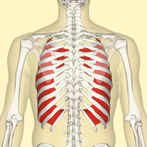
The diaphragm is a sheet of muscles beneath your thoracic basket or rib cage (Figure 11.4 and 11.5). The external and intercostalis are muscles situated in between your costae or ribs (Figure 11.6 and 11.7). Together, the three sets of muscles assist us to breathe in (inspiration) and breathe out (expiration) during alveolar respiration. They act to elevate and expand the rib cage and thus the lungs become elevated. During expiration, the rib cage becomes reduced in size.


Muscles of the Abdomen
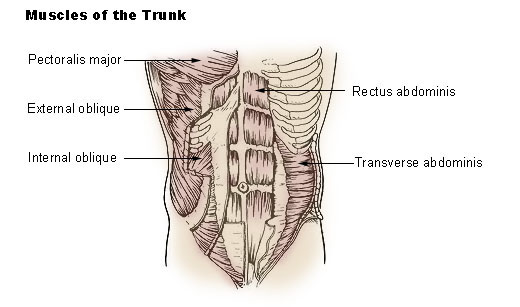
The muscles that cover the abdominal cavity are the abdominal muscles (Figure 11.8 and 11.9). Located between the ribs and the pelvis, they are sheets of flat muscles and are prominent. The muscles are the external obliques, the internal obliques, the transversus abdominis, and the rectus abdominis. The external obliques are situated on the lateral sides of the abdomen. The paired rectus abdominis run vertically from pelvis to the xiphoid process and ribs. The internal obliques runs laterally. The transversus abdominis muscle is deep to the internal oblique muscle.

Pectoral Muscles
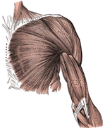
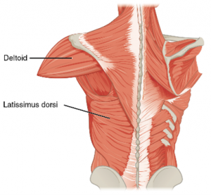
The pectoral muscles (Figure 11.10, 11.11 and 11.12) are attached to the pectoral girdle. The pectoralis major is the set of fan-shaped prominent muscles on the chest. Its origin is in the clavicle, sternum and abdomen. It inserts in the tubercle of the humerus bone. The pectoralis minor is deep to the pectoralis major. Its origin is the 3rd to 5th rib. The insertion is in coracoid process of the scapula. The serratus anterior muscle originates in the first to 8th rib. It inserts in the scapula. The trapezius is a large muscle originating in the C7 to T12 vertebrae. Its insertion is in the clavicle and scapula. The rhomboid muscle is broad and originates in the thoracic vertebrae T2-T5. It inserts in the scapula. The latissimus dorsi is a broad, flat muscle. It has several points of origin: T7-T12, thoracolumbar fascia, iliac crest and scapula. Its insertion is in the humerus. The teres major originates in the lateral border of the scapula and inserts in the humerus.

Example of Lower Limb Muscles – Gluteus Maximus, Sartorius, Hamstring Muscles
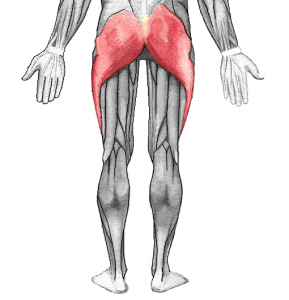
The gluteus maximus (Figure 11.13) is the largest, fleshy muscle dorsally. It extends from the hips (sacrum, ileum and fascia) to the femur. The gluteus minimus is a triangular shaped muscle located deep to the gluteus maximus. It originates in the ilium and inserts in the greater tuberosity of the femur. Gluteus medius originates in the ileum and inserts in the greater trochanter of the femur. Review the skeleton bones.
Sartorius
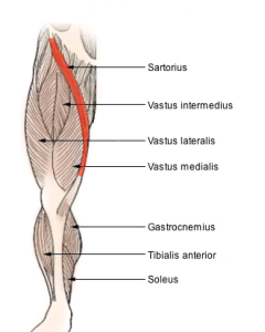
The sartorius muscle (Figure 11.14) is a belt-like muscle that extends diagonally from the anterior superior iliac spine to the proximal end of tibia below medial condyle. It crosses from hip to knee joints. The origin is in the iliac bone and insertion in the tibia. It is the longest muscle in the body. The muscle is referred to as a tailor’s muscle because they used to sit cross-legged to sew on a sewing machine.
Hamstring Muscles
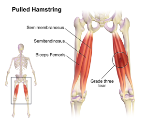
The hamstring muscles are a set of three muscles (Figure 11.15): semi-tendinosus, biceps femoris and semi-membranosus. They all have their origin in the ischium. The insertion is in the tibia and fibula. The hamstrings connect bones of the pelvis, knees and lower leg. The biceps femoris is long. It starts in the thigh and attaches to the fibula. It is laterally situated. The semimembranosus begins at the pelvis and ends in the tibia. The semitendinosus lies between the semimembranosus and biceps femoris. It is the longest of the three muscles. The hamstring muscles are an important set of muscles if only because sports people injure them.
Pre-Laboratory Questions
- Name the main facial muscles. State the origin and insertion of each.
- Name the main thoracic muscles. What are their functions?
- List muscles of the abdominal wall. What are their functions?
- List the main muscles of the pectoral girdle. What are their points of origin and insertion? What is their function?
- List the main muscles of the upper limb.
- List the main muscles of the lower limb. What are their points of origin and insertion? What are their functions.
Exercises
- Exercise 1 Name and identify facial muscles
- Exercise 2 Identification of thoracic muscles
- Exercise 3 Identification of muscles of the abdomen
- Exercise 4 Identification of pectoral muscles
- Exercise 5 Identification of muscles of lower limb
Exercise 1. Name and identify facial muscles
Required Materials
- Human Muscle Model (life size)
- Male Muscular Figure
- Mini Human Muscular Figure
- The Muscular System Poster
- Post-it notes
Procedure
1. Feel these muscles on your face (Figure 11.3): buccinators, mentalis, orbicularis oris, orbicularis oculi, platysma, zygomaticus. Ask your instructor to show the model on the table that displays muscles. Use the model to identify each muscle. Blow up your mouth as if you were blowing up a balloon. The cheek muscles are the buccinators.
2. Mentalis muscle is the lower lip muscle. Feel it. Now, use your lower lip to pout. The mentalis muscle helps you to pout. Identify the mentalis muscle on the model on demonstration.
3. Orbicularis oris is the set of muscles around your mouth. Protrude your lips as if you are kissing. Orbicularis muscles help you to do that. Purse your lips. “Orb” means “globe”, “round”. “Oris” means “opening”.
4. Run your fingers around your eyelid. The muscle you will feel is the orbicularis oculi muscle. It helps you to close your eyes. “Oculus” means a round window.
5. Platysma is a flat sheet of muscles extending from below the chin to the top of the throat. You can feel this muscle if you stretch your chin to look up at the ceiling.
6. Zygomaticus muscle is attached to the zygomaticus bone. It forms an angle with the upper lip. Together with the buccinator, it helps to form speech and facial expressions. Smile or laugh….and it is these set of muscles that will go into action.
7. Label the muscles you found on the muscle model using post-it notes. Take a picture. Alternatively, you can draw and label these muscles below.

8. Make a chart showing functions of the buccinators, orbicularis oris, orbicularis oculi, platysma and zygomaticus.
| Muscle Type | Function |
|---|---|
| Buccinators | |
| Orbicularis oris | |
| Orbicularis sculi | |
| Platysma | |
| Zygomaticus |
Exercise 2. Identification of thoracic muscles
Required Materials
- Human Muscle Model (life size)
- Male Muscular Figure
- Mini Human Muscular Figure
- The Muscular System Poster
- Post-it notes
Procedure
- Run your fingers down to the end of the thoracic basket and tuck them in to feel the diaphragm (Figure 11.4 and 11.5). it is the muscle that separates your thoracic cavity from your abdominal cavity. Identify it on the model on demonstration. Ask your instructor to point it out to you.
- The intercostal muscles are located in-between the ribs (Figure 11.6 and 11.7). Put your fingers to the sides of your thoracic basket also to feel the external intercostal muscles. The intercostal muscles are deep to the external intercostals.
- Use the post-it notes to label these muscles on the models. Take a picture and insert it below. Alternatively, you can draw and label these muscles on your drawing.
Exercise 3. Identification of muscles of the abdomen
Required Materials
- Human Muscle Model (life size)
- Male Muscular Figure
- Mini Human Muscular Figure
- The Muscular System Poster
- Post-it notes
Procedure
- Feel the muscles covering the abdomen (Figure 11.8 and 11.9). You may not be able to identify them. But use the model to study the four muscles: external oblique, internal oblique, rectus abdominis and transverse abdominis. To locate the transversus abdominis muscle, feel the side of your abdomen just where the pelvic bone is located. Contract the muscle and hold for 5-6 seconds. The rectus abdominis can be felt by running the fingers from below the xiphoid process to the pelvis.
- Make a chart showing the origin and insertion of the external obliques, the internal obliques, the transversus abdominis, and the rectus abdominis (Hint: Consider the sternum, ribs, pubic bones).
| Origin | Insertion | |
| external obliques | ||
| internal obliques | ||
| transverse abdominis | ||
| rectus abdominis |
3. Use the post-it notes to label these muscles on the models. Take a picture and insert below. Alternatively, you can draw and label these muscles below.
Exercise 4. Identification of pectoral muscles
Required Materials
- Human Muscle Model (life size)
- Male Muscular Figure
- Mini Human Muscular Figure
- The Muscular System Poster
- Post-it notes
Procedure
- The pectoralis major muscle is best seen in the model on demonstration (Figure 11.2). Study its origin and insertion. The pectoralis minor and serratus major can also be viewed on the model.
- The trapezius, rhomboid (Figure 11.12) and latissimus dorsi (Figure 11.11) can best be felt by placing your hands on the dorsal / posterior aspect of your body.
- Use the post-it notes to label these muscles on the model. Take a picture and insert it below. Alternatively, you can draw and label these muscles below.
Exercise 5. Identification of muscles of the lower limb
Required Materials
- Human Muscle Model (life size)
- Male Muscular Figure
- Mini Human Muscular Figure
- The Muscular System Poster
- Post-it notes
Procedure
- You can feel the gluteus muscle with your hands. The gluteus maximus muscle is your buttock (Figure 11.13).
- With your hands, feel the ileum, sacrum and coccyx of your pelvic bones. These are the three bones where the gluteus maximus muscle originates. They insert on the tuberosity of the femur which you can also feel. Now, trace the muscle on the demonstration model.
- For the sartorius muscle (Figure 11.14), You can sit cross-legged on the floor. Or, you can cross one leg over the other sitting on a stool. Using your thumbs, feel the width of the sartorius muscle in your inner thigh. Now run the two thumbs along the edges of the muscle to the knee/ hips. By doing this, you will be feeling the sartorius muscle from the hip to the tibia.
- The hamstring muscles (Figure 11.15) These can be felt easily by your fingers and hand. Feel them by placing your hands to the back of your thighs. They run along the length of the femur.
- Use the post-it notes to label these muscles on the model. Take a picture and insert it below. Alternatively, you can draw and label these muscles below. For gluteus maximus and hamstrings, use the posterior view. For sartorius, use the anterior view.
Posterior view of lower limb muscles:
Anterior view of lower limb muscles:
Post-laboratory Questions
- What is the common name for the buccinator muscles?
- What are the muscles that form your upper and lower lips, eyes?
- If a male person was shaving the underside of his maxillary region by stretching it and looking upwards, what is the muscle he would be feeling?
- State the functions of the (a) latissimus dorsi and (b) trapezius muscles.
- Name the origin of the pectoralis major muscle.
- What is the tailor’s muscle? Why is it so called?
- Where do the hamstring muscles originate and insert?
