
9 Chapter 9 Joints
By Ganesan L. Kamatchi
Motivation.
Osteoarthritis is a type of joint disease that results from breakdown of joint cartilage and underlying bone. The most common symptoms are joint pain and stiffness. Usually, the symptoms progress slowly over years. Initially they may occur only after exercise but can become constant over time. Other symptoms may include joint swelling, decreased range of motion, and, when the back is affected, weakness or numbness of the arms and legs. The most commonly involved joints are the two near the ends of the fingers and the joint at the base of the thumbs; the knee and hip joints; and the joints of the neck and lower back. Joints on one side of the body are often more affected than those on the other. The symptoms can interfere with work and normal daily activities. Unlike some other types of arthritis, only the joints, not internal organs, are affected. Osteoarthritis is the most common form of arthritis, affecting about 237 million people, or 3.3% of the world’s population. In the United States, 30 to 53 million people are affected. It becomes more common as people become older.
Learning Objectives
Upon completion of the work in this chapter students should be able to:
- Define and classify articulations
- Classify fibrous joints with examples
- Describe and classify cartilaginous joints
- Describe and classify synovial joints
- Describe the types of movements possible in synovial joints
Background.
The adult human body has 206 bones, and with the exception of the hyoid bone in the neck, each bone is connected to at least one other bone. Joints are the location where bones come together. Many joints allow for movement between the bones. At these joints, the articulating surfaces of the adjacent bones can move smoothly against each other. However, the bones of other joints may be joined to each other by connective tissue or cartilage. These joints are designed for stability and provide for little or no movement. Importantly, joint stability and movement are related to each other. This means that stable joints allow for little or no mobility between the adjacent bones. Conversely, joints that provide the most movement between bones are the least stable. Understanding the relationship between joint structure and function will help to explain why particular types of joints are found in certain areas of the body.
The articulating surfaces of bones at stable types of joints, with little or no mobility, are strongly united to each other. For example, most of the joints of the skull are held together by fibrous connective tissue and do not allow for movement between the adjacent bones. This lack of mobility is important because the skull bones serve to protect the brain. Similarly, other joints united by fibrous connective tissue allow for very little movement, which provides stability and weight-bearing support for the body. For example, the tibia and fibula of the leg are tightly united to give stability to the body when standing. At other joints, the bones are held together by cartilage, which permits limited movements between the bones. Thus, the joints of the vertebral column only allow for small movements between adjacent vertebrae, but when added together, these movements provide the flexibility that allows your body to twist, or bend to the front, back, or side. In contrast, at joints that allow for wide ranges of motion, the articulating surfaces of the bones are not directly united to each other. Instead, these surfaces are enclosed within a space filled with lubricating fluid, which allows the bones to move smoothly against each other. These joints provide greater mobility, but since the bones are free to move in relation to each other, the joint is less stable. Most of the joints between the bones of the appendicular skeleton are this freely moveable type of joint. These joints allow the muscles of the body to pull on a bone and thereby produce movement of that body region. Your ability to kick a soccer ball, pick up a fork, and dance the tango depend on mobility at these types of joints.
Fibrous Joints
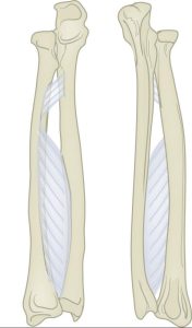
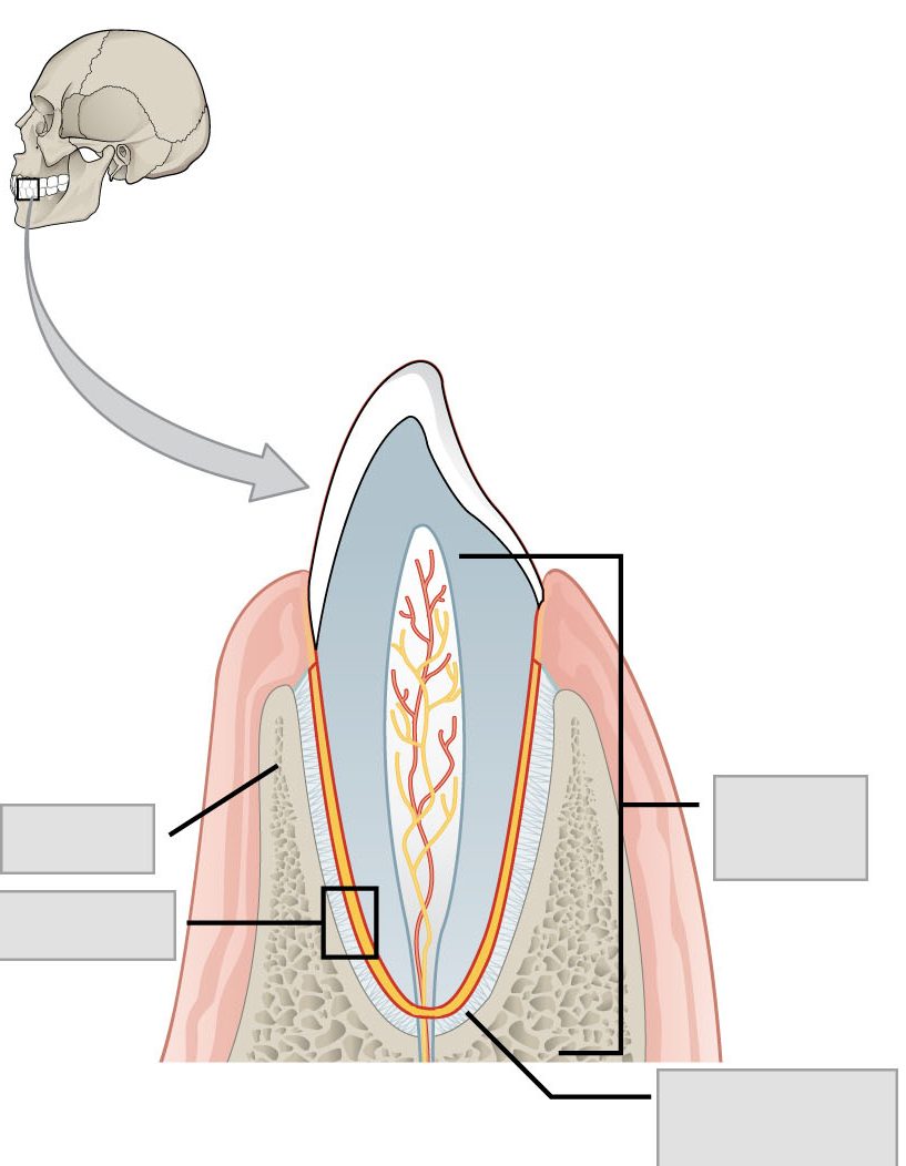
At a fibrous joint, the adjacent bones are directly connected to each other by fibrous connective tissue, and thus the bones do not have a joint cavity between them (Figure 9.2 a-c). The gap between the bones may be narrow or wide. There are three types of fibrous joints. A suture is the narrow fibrous joint found between most bones of the skull. At a syndesmosis joint, the bones are more widely separated but are held together by a narrow band of fibrous connective tissue called a ligament or a wide sheet of connective tissue called an interosseous membrane. This type of fibrous joint is found between the shaft regions of the long bones in the forearm and in the leg. Lastly, a gomphosis is the narrow fibrous joint between the roots of a tooth and the bony socket in the jaw into which the tooth fits.
Cartilagenous Joints
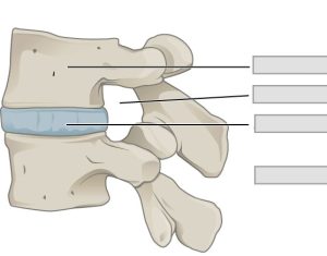
As the name indicates, at a cartilaginous joint, the adjacent bones are united by cartilage, a tough but flexible type of connective tissue. These types of joints lack a joint cavity and involve bones that are joined together by either hyaline cartilage or fibrocartilage (Figure 9.3 a-b). There are two types of cartilaginous joints. A synchondrosis is a cartilaginous joint where the bones are joined by hyaline cartilage. Also classified as a synchondrosis are places where bone is united to a cartilage structure, such as between the anterior end of a rib and the costal cartilage of the thoracic cage, or in the epiphyseal plate of a growing long bone. The second type of cartilaginous joint is a symphysis, where the bones are joined by fibrocartilage. The pubic portions of the right and left hip bones of the pelvis are joined together by fibrocartilage, forming the pubic symphysis. An intervertebral disc unites the bodies of adjacent vertebrae within the vertebral column. Intervertebral discs are made of fibrocartilage and thereby structurally form a symphysis type of cartilaginous joint.
Cartilaginous joints are also functionally classified as either a synarthrosis or an amphiarthrosis joint.
Synovial Joints
Synovial joints are the most common type of joint in the body (Figure 9.4). A key structural characteristic for a synovial joint that is not seen at fibrous or cartilaginous joints is the presence of a joint cavity. This fluid-filled space is the site at which the articulating surfaces of the bones contact each other. Also unlike fibrous or cartilaginous joints, the articulating bone surfaces at a synovial joint are not directly connected to each other with fibrous connective tissue or cartilage. This gives the bones of a synovial joint the ability to move smoothly against each other, allowing for increased joint mobility.
Synovial joints are characterized by the presence of a joint cavity. The walls of this space are formed by the articular capsule, a fibrous connective tissue structure that is attached to each bone just outside the area of the bone’s articulating surface. The bones of the joint articulate with each other within the joint cavity.
Friction between the bones at a synovial joint is prevented by the presence of the articular cartilage, a thin layer of hyaline cartilage that covers the entire articulating surface of each bone. However, unlike at a cartilaginous joint, the articular cartilages of each bone are not continuous with each other. Instead, the articular cartilage acts like a Teflon® coating over the bone surface, allowing the articulating bones to move smoothly against each other without damaging the underlying bone tissue. Lining the inner surface of the articular capsule is a thin synovial membrane. The cells of this membrane secrete synovial fluid (synovia = “a thick fluid”), a thick, slimy fluid that provides lubrication to further reduce friction between the bones of the joint. This fluid also provides nourishment to the articular cartilage, which does not contain blood vessels. The ability of the bones to move smoothly against each other within the joint cavity, and the freedom of joint movement this provides, means that each synovial joint is functionally classified as a diarthrosis.
Outside of their articulating surfaces, the bones are connected together by ligaments, which are strong bands of fibrous connective tissue. These strengthen and support the joint by anchoring the bones together and preventing their separation. Ligaments allow for normal movements at a joint, but limit the range of these motions, thus preventing excessive or abnormal joint movements. Ligaments are classified based on their relationship to the fibrous articular capsule. An extrinsic ligament is located outside of the articular capsule, an intrinsic ligament is fused to or incorporated into the wall of the articular capsule, and an intracapsular ligament is located inside of the articular capsule.
At many synovial joints, additional support is provided by the muscles and their tendons that act across the joint. A tendon is the dense connective tissue structure that attaches a muscle to bone. As forces acting on a joint increase, the body will automatically increase the overall strength of contraction of the muscles crossing that joint, thus allowing the muscle and its tendon to serve as a “dynamic ligament” to resist forces and support the joint. This type of indirect support by muscles is very important at the shoulder joint, for example, where the ligaments are relatively weak.
A few synovial joints of the body have a fibrocartilage structure located between the articulating bones. This is called an articular disc, which is generally small and oval-shaped, or a meniscus, which is larger and C-shaped. These structures can serve several functions, depending on the specific joint. In some places, an articular disc may act to strongly unite the bones of the joint to each other. Examples of this include the articular discs found at the sternoclavicular joint or between the distal ends of the radius and ulna bones. At other synovial joints, the disc can provide shock absorption and cushioning between the bones, which is the function of each meniscus within the knee joint. Finally, an articular disc can serve to smooth the movements between the articulating bones, as seen at the temporomandibular joint. Some synovial joints also have a fat pad, which can serve as a cushion between the bones.
Additional structures located outside of a synovial joint serve to prevent friction between the bones of the joint and the overlying muscle tendons or skin. A bursa (plural = bursae) is a thin connective tissue sac filled with lubricating liquid. They are located in regions where skin, ligaments, muscles, or muscle tendons can rub against each other, usually near a body joint (Figure 9.5).
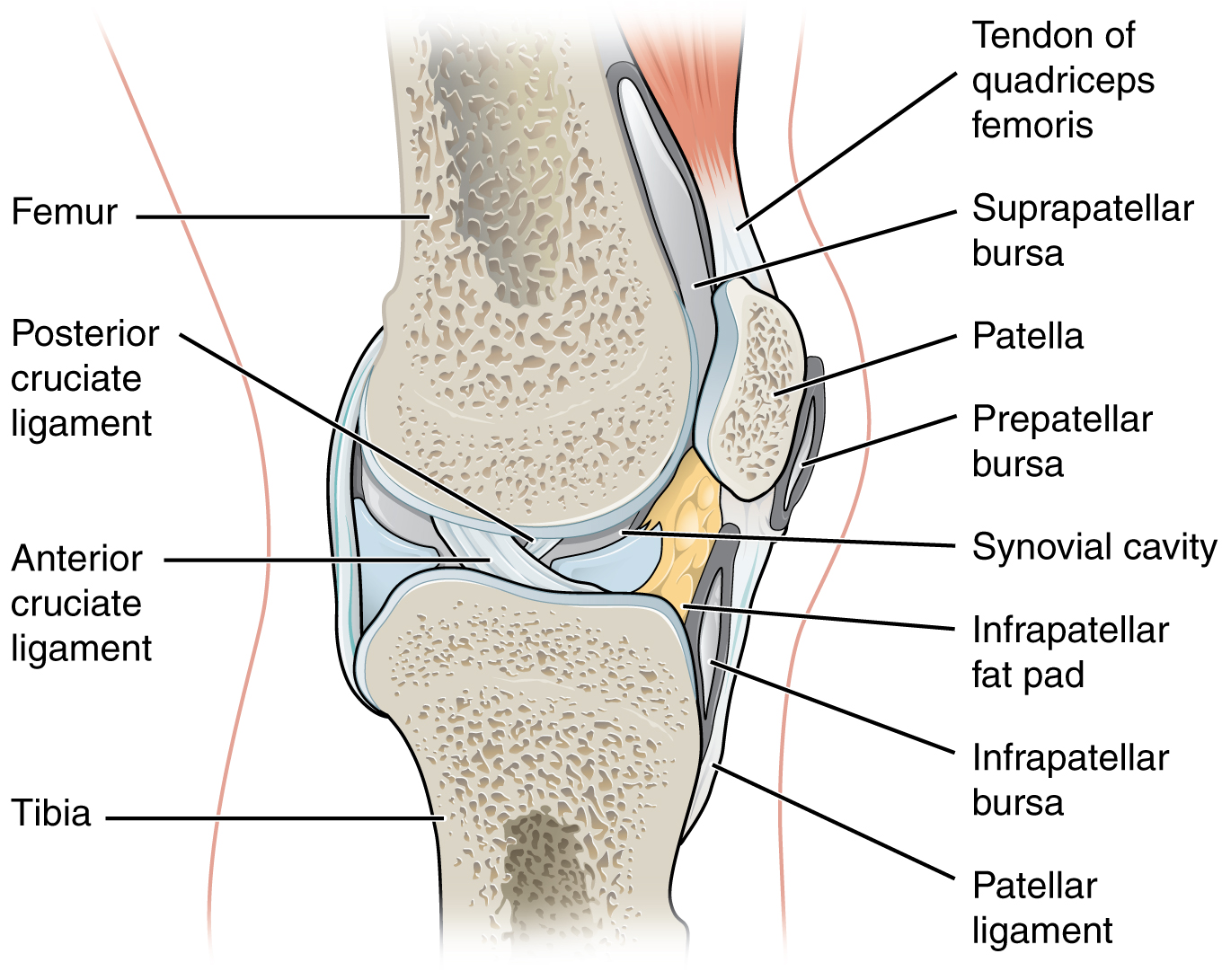
Bursae reduce friction by separating the adjacent structures, preventing them from rubbing directly against each other. Bursae are classified by their location. A subcutaneous bursa is located between the skin and an underlying bone. It allows skin to move smoothly over the bone. Examples include the prepatellar bursa located over the kneecap and the olecranon bursa at the tip of the elbow. A submuscular bursa is found between a muscle and an underlying bone, or between adjacent muscles. These prevent rubbing of the muscle during movements. A large submuscular bursa, the trochanteric bursa, is found at the lateral hip, between the greater trochanter of the femur and the overlying gluteus maximus muscle. A subtendinous bursa is found between a tendon and a bone. Examples include the subacromial bursa that protects the tendon of shoulder muscle as it passes under the acromion of the scapula, and the suprapatellar bursa that separates the tendon of the large anterior thigh muscle from the distal femur just above the knee.
A tendon sheath is similar in structure to a bursa, but smaller. It is a connective tissue sac that surrounds a muscle tendon at places where the tendon crosses a joint. It contains a lubricating fluid that allows for smooth motions of the tendon during muscle contraction and joint movements.
Classification of Synovial Joints
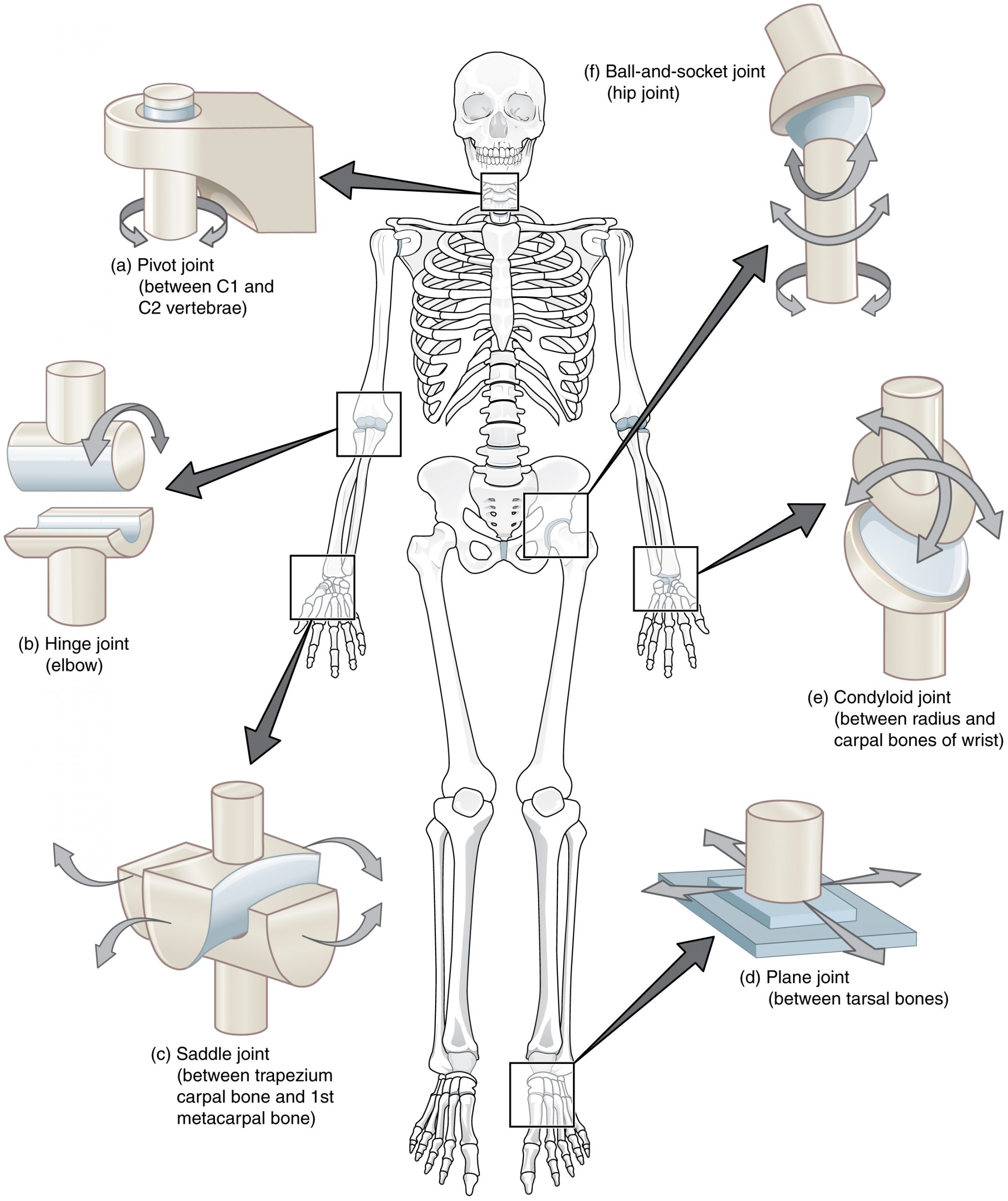
Synovial joints are subdivided based on the shapes of the articulating surfaces of the bones that form each joint. The six types of synovial joints are pivot, hinge, condyloid, saddle, plane, and ball-and socket-joints (Figure 9.6).
Pivot Joint
At a pivot joint, a rounded portion of a bone is enclosed within a ring formed partially by the articulation with another bone and partially by a ligament (see Figure 9.6a). The bone rotates within this ring. Since the rotation is around a single axis, pivot joints are functionally classified as a uniaxial diarthrosis type of joint. An example of a pivot joint is the atlantoaxial joint, found between the C1 (atlas) and C2 (axis) vertebrae. Here, the upward projecting dens of the axis articulates with the inner aspect of the atlas, where it is held in place by a ligament. Rotation at this joint allows you to turn your head from side to side. A second pivot joint is found at the proximal radioulnar joint. Here, the head of the radius is largely encircled by a ligament that holds it in place as it articulates with the radial notch of the ulna. Rotation of the radius allows for forearm movements.
Hinge Joint
In a hinge joint, the convex end of one bone articulates with the concave end of the adjoining bone (see Figure 9.6b). This type of joint allows only for bending and straightening motions along a single axis, and thus hinge joints are functionally classified as uniaxial joints. A good example is the elbow joint, with the articulation between the trochlea of the humerus and the trochlear notch of the ulna. Other hinge joints of the body include the knee, ankle, and interphalangeal joints between the phalanx bones of the fingers and toes.
Saddle Joint
At a saddle joint, both of the articulating surfaces for the bones have a saddle shape, which is concave in one direction and convex in the other (see Figure 9.6c). This allows the two bones to fit together like a rider sitting on a saddle. Saddle joints are functionally classified as biaxial joints. The primary example is the first carpometacarpal joint, between the trapezium (a carpal bone) and the first metacarpal bone at the base of the thumb. This joint provides the thumb the ability to move away from the palm of the hand along two planes. Thus, the thumb can move within the same plane as the palm of the hand, or it can jut out anteriorly, perpendicular to the palm. This movement of the first carpometacarpal joint is what gives humans their distinctive “opposable” thumbs. The sternoclavicular joint is also classified as a saddle joint.
Plane Joint
At a plane joint (gliding joint), the articulating surfaces of the bones are flat or slightly curved and of approximately the same size, which allows the bones to slide against each other (see Figure 9.6d). The motion at this type of joint is usually small and tightly constrained by surrounding ligaments. Based only on their shape, plane joints can allow multiple movements, including rotation. Thus plane joints can be functionally classified as a multiaxial joint. However, not all of these movements are available to every plane joint due to limitations placed on it by ligaments or neighboring bones. Thus, depending upon the specific joint of the body, a plane joint may exhibit only a single type of movement or several movements. Plane joints are found between the carpal bones (intercarpal joints) of the wrist or tarsal bones (intertarsal joints) of the foot, between the clavicle and acromion of the scapula (acromioclavicular joint), and between the superior and inferior articular processes of adjacent vertebrae (zygapophysial joints).
Condyloid Joint
At a condyloid joint (ellipsoid joint), the shallow depression at the end of one bone articulates with a rounded structure from an adjacent bone or bones (see Figure 9.6e). The knuckle (metacarpophalangeal) joints of the hand between the distal end of a metacarpal bone and the proximal phalanx bone are condyloid joints. Another example is the radiocarpal joint of the wrist, between the shallow depression at the distal end of the radius bone and the rounded scaphoid, lunate, and triquetrum carpal bones. In this case, the articulation area has a more oval (elliptical) shape. Functionally, condyloid joints are biaxial joints that allow for two planes of movement. One movement involves the bending and straightening of the fingers or the anterior-posterior movements of the hand. The second movement is a side-to-side movement, which allows you to spread your fingers apart and bring them together, or to move your hand in a medial-going or lateral-going direction.
Ball-and-Socket Joint
The joint with the greatest range of motion is the ball-and-socket joint. At these joints, the rounded head of one bone (the ball) fits into the concave articulation (the socket) of the adjacent bone (see Figure 9.6f). The hip joint and the glenohumeral (shoulder) joint are the only ball-and-socket joints of the body. At the hip joint, the head of the femur articulates with the acetabulum of the hip bone, and at the shoulder joint, the head of the humerus articulates with the glenoid cavity of the scapula.
Ball-and-socket joints are classified functionally as multiaxial joints. The femur and the humerus are able to move in both anterior-posterior and medial-lateral directions and they can also rotate around their long axis. The shallow socket formed by the glenoid cavity allows the shoulder joint an extensive range of motion. In contrast, the deep socket of the acetabulum and the strong supporting ligaments of the hip joint serve to constrain movements of the femur, reflecting the need for stability and weight-bearing ability at the hip.
Pre-Laboratory Questions
After you complete reviewing the Background information provided, please answer the following questions prior to doing the lab exercises.
- What is the functional classification of joints?
A) synarthroses.
B) amphiarthroses.
C) diarthroses.
D) all of the above.
- What is the structural classification of joints?
A) fibrous joint.
B) cartilaginous joint.
C) synovial joint.
D) all of the above.
- What is the most common type of joint in the human body?
A) fibrous joints.
B) cartilaginous joints.
C) synovial joints.
- Fibrous joints belong to which functional type of joints?
A) synarthroses.
B) amphiarthroses.
C) diarthroses.
- What is the only movable bone in the human skull?
A) mandible.
B) frontal.
C) parietal.
D) occipital.
Exercises
- Exercise 1 Fibrous Joints
- Exercise 2 Cartilagenous Joints
- Exercise 3 Synovial Joints
Exercise 1 Fibrous Joints
Exercise 1a. Suture
Required Materials
- Skull models
- Poster of the skeletal system
Procedure
- Examine a skull model and study the suture joints between the bones of the skull (Figure 9.2a).
- Identify the structures in the list below and label these on the figure given.
· Frontal bone · Name of suture 3 · Name of suture 1 · Parietal bone · Name of suture 2 · Occipital bone
Exercise 1b. Syndesmosis
Required Materials
- Articulated skeleton model
- Poster of the skeletal system
Procedures
- Examine the whole skeleton and observe the joint between the radius and ulna and its parts from all sides (Figure 9.2b)
- Identify the structures in the list below and label these on the figure given.
· Antebrachial interosseous membrane · Ulna · Radius
Exercise 1c. Gomphosis
Required Materials
- Tooth in the human digestive system model
- Model of Half of the Human Head
- Median Section of the Head Model
- Skull model
- Poster of the skeletal system
Procedure
- Examine a model of gomphosis joint and observe its parts from all sides (Figure 9.2c)
- Identify the structures in the list below and label them in the figure given.
· Alveolar process of mandible · Periodontal ligament · Gomphosis · Root of tooth
Exercise 2 Cartilaginous Joints
Exercise 2a. Synchondrosis
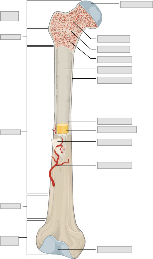
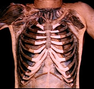
In a long bone, the hyaline cartilage of the epiphyseal plate (growth plate) forms a synchondrosis that unites the shaft (diaphysis) and end (epiphysis) of the bone and allows the bone to grow in length. The costochondral joint between the first rib and the manubrium of the sternum also forms a synchondrosis.
Required Materials
- Rib cage on articulated skeleton
- Cut long bones on bone tissue model
- Poster of the skeletal system
Procedure
- Examine a longitudinal section of a long bone and the rib cage and study the synchondrosis joint.
- Identify the structures in the list below and label these on the figures given.
· Body · First rib · Clavicle · Manubrium · Diaphysis · Clavicle · Epiphyseal plate · Xiphoid process · Epiphysis
Exercise 2b. Symphysis
Required Materials
- Flexible vertebrate column model (with pelvic girdle)
- Poster of the skeletal system
Procedure
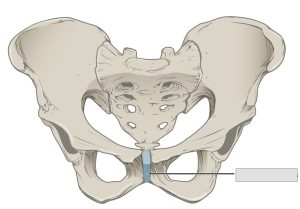
- Examine a pelvic girdle and vertebral column and study the symphysis joint (Figure 9.3).
- Identify the structures in the list below and label them on the figures given.
· Acetabulum · Ischium · Coccyx · Pubic symphysis · Iliac crest · Pubis · Ilium · Sacrum · Intervertebral disc · Vertebral body
Exercise 3 Synovial Joints
Required Materials
- Miniature functional joints model set
- Articulated skeleton
- Skeletal system poster
Procedure
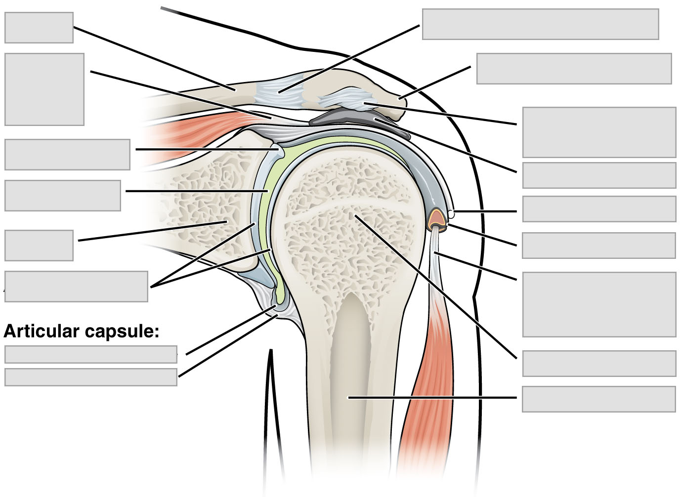
- Examine a model of the shoulder joint or the shoulder joint of a skeleton and study the structure of ball-and-socket joint (Figure 4, 5, and 6f).
- Identify the structures in the list below and label these in the figure given.
· Acromioclavicular joint · Glenoid labrum · Acromion of scapula · Head of humerus · Articular capsule – synovial membrane · Humerus · Articular capsule – fibrous membrane · Tendon of supraspinatus muscle · Articular capsule · Scapula · Articular cartilage · Subacromial bursa · Clavicle · Tendon of biceps brachii muscles (long head) · Coracoacromial ligament · Tendon sheath · Glenoid cavity
Post-laboratory Questions
- A synarthrosis is
A) always made of cartilage.
B) a joint that has a capsule.
C) a joint within a fetus that ossifies during early development.
D) immobile.
E) slightly mobile.
- Which of the following joints contain an interosseous membrane?
A) gomphosis.
B) suture.
C) syndesmosis.
- A synarthrotic joint would have
A) high mobility and high stability.
B) high mobility and low stability.
C) low mobility and low stability.
D) low mobility and high stability.
- Functionally, a gomphosis is categorized as a
A) cartilagenous joint.
B) diarthrosis.
C) synarthrosis.
D) synovial joint.
- In fibrous joints, the articulating surfaces are held together by
A) dense regular connective tissue.
B) areolar connective tissue.
C) dense irregular
D) fibrocartilage.
E) articular cartilage.
6. Structurally, a syndesmosis is a _________ joint; functionally, it is a _________
A) cartilagenous; diarthrosis
B) cartilagenous; amphiarthrosis
C) fibrous; amphiarthrosis
D) fibrous; synarthrosis
E) ball and socket; pivot
- Sutures are joints that are found
A) throughout the axial and appendicular skeletons.
B) between all bones and teeth of the skull.
C) between certain bones of the skull.
D) only where a facial bone articulates with a cranial bone.
- The interosseous membrane between the radius and the ulna is an example of a
A) synchondrosis.
B) suture.
C) synostosis.
D) synarthrosis.
E) syndesmosis.
- The pubic symphysis is classified as a
A) cartilaginous joint and an amphiarthrosis.
B) fibrous joint and a synarthrosis.
C) synovial joint and a diarthrosis.
D) cartilaginous joint and a synarthrosis.
E) fibrous joint and an amphiarthrosis.
- What type of cartilage is located between the bones in a symphysis?
A) elastic cartilage
B) reticular cartilage
C) hyaline cartilage
D) fibrocartilage
- What type of cartilage do the intervertebral discs belong to?
A) elastic cartilage
B) reticular cartilage
C) hyaline cartilage
D) fibrocartilage
- What type of cartilage do the costochondral cartilages belong to?
A) elastic cartilage
B) reticular cartilage
C) hyaline cartilage
D) fibrocartilage
- An articular capsule is present in
A) fibrous joints.
B) fibrous joints and cartilaginous joints.
C) synovial joints.
D) fibrous joints and synovial joints.
E) all joints.
- Fluid-filled sacs that cushion synovial joints are called
A) fat pads.
B) articular discs.
C) bursae.
D) menisci.
E) diarthroses.
- Synovial fluid is
A) a watery fluid produced by capsular ligaments.
B) an oily fluid produced by the synovial membrane.
C) a watery fluid produced by hyaline cartilage.
D) an oily fluid produced by articular cartilage.
- The largest and most complex synovial joint is the
A) mandibular joint
B) carpal joint
C) elbow joint
D) knee joint
E) finger joints
- Which joint is the most easily dislocated?
A) mandibular joint.
B) carpal joint.
C) shoulder joint.
D) knee joint.
E) hip joint.
