
8 Chapter 8 The Appendicular Skeleton
By Ganesan L. Kamatchi
Motivation.

Gorham-Stout disease is a rare and enigmatic condition involving various skeletal locations. It is also called massive bone disease, vanishing bone disease, phantom bone disease, massive osteolysis, Gorham–Stout syndrome and Gorham’s disease. It is characterized by destruction of osseous matrix and proliferation of vascular structures with benign origin. Despite the extensive investigation of the pathogenetic mechanisms of the disease, its etiology of GSD is poorly understood and the cause of excessive bone resorption in GSD patients has not been clarified. Several studies proposed that the abnormal proliferation of endothelial-lined vessels could promote bone resorption. Bone normally do not have lymphatic vessels and the presence of lymphatics in GSD patients have been confirmed histologically and biochemically. Gorham-Stout disease can affect any part of the skeleton, but the pelvis, long bones, and shoulder girdles are the most frequently involved [Kaissi et al., Medicines (Basel) 6: 54, 2019].
Learning Objectives
Upon completion of the work in this chapter students should be able to:
- Identify and label the pectoral girdle bone structures in the clavicle and scapula samples provided
- Identify the bones of the upper limbs and label their structures
- Label the bones and structures within the pelvic girdle after identifying on an articulated skeleton
- Identify the bones of the lower limbs and label these and their associated structures
Background.
Your skeleton provides the internal supporting structure of the body. The adult axial skeleton consists of 80 bones that form the head and body trunk. Attached to this are the limbs, whose 126 bones constitute the appendicular skeleton (Figure 8.2). These bones are divided into two groups: the bones that are located within the limbs themselves, and the girdle bones that attach the limbs to the axial skeleton. The bones of the shoulder region form the pectoral girdle, which anchors the upper limb to the thoracic cage of the axial skeleton. The lower limb is attached to the vertebral column by the pelvic girdle.
Because of our upright stance, different functional demands are placed upon the upper and lower limbs. Thus, the bones of the lower limbs are adapted for weight-bearing support and stability, as well as for body locomotion via walking or running. In contrast, our upper limbs are not required for these functions. Instead, our upper limbs are highly mobile and can be utilized for a wide variety of activities. The large range of upper limb movements, coupled with the ability to easily manipulate objects with our hands and opposable thumbs, has allowed humans to construct the modern world in which we live.

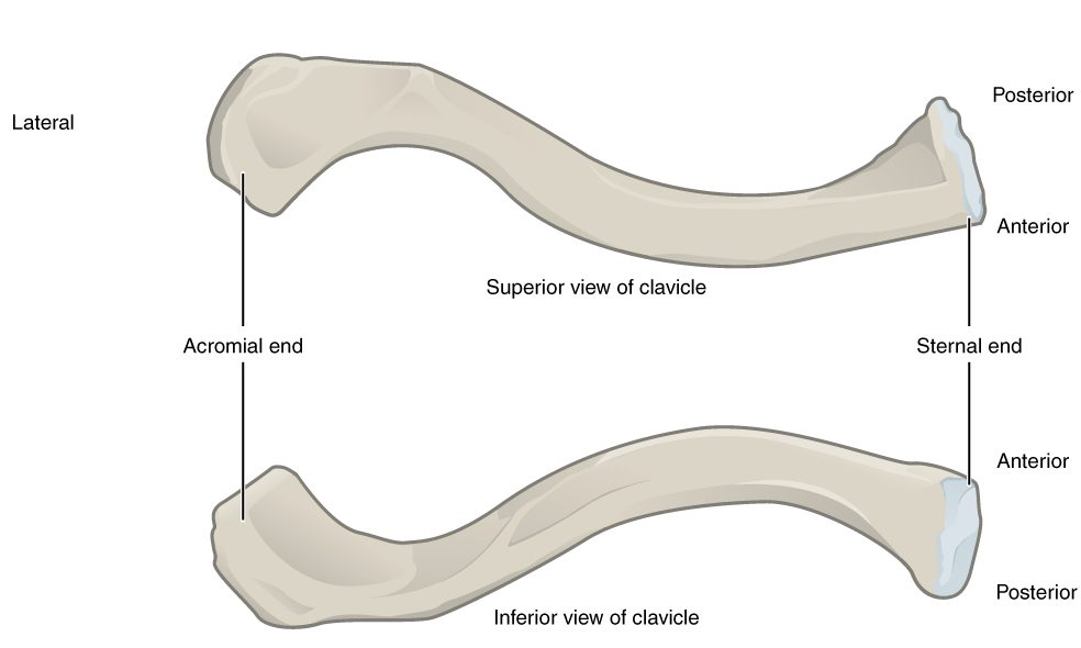
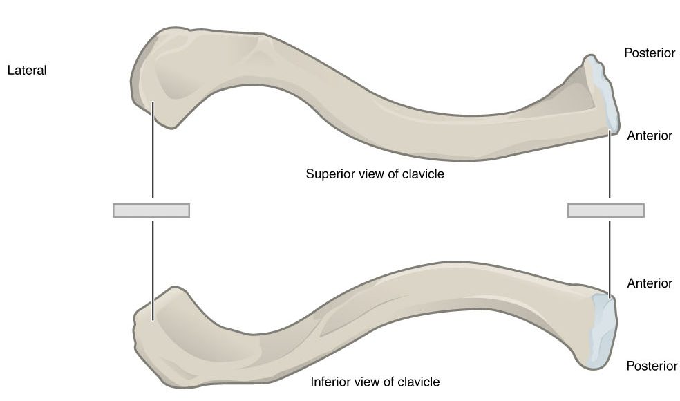
The Clavicle
The clavicle is the only long bone that lies in a horizontal position in the body (see Figure 8.3). The clavicle has several important functions. First, anchored by muscles from above, it serves as a strut that extends laterally to support the scapula. This in turn holds the shoulder joint superiorly and laterally from the body trunk, allowing for maximal freedom of motion for the upper limb. The clavicle also transmits forces acting on the upper limb to the sternum and axial skeleton. Finally, it serves to protect the underlying nerves and blood vessels as they pass between the trunk of the body and the upper limb.
The Scapula
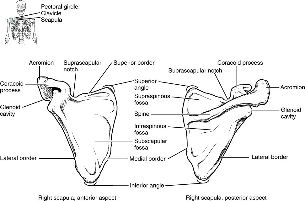
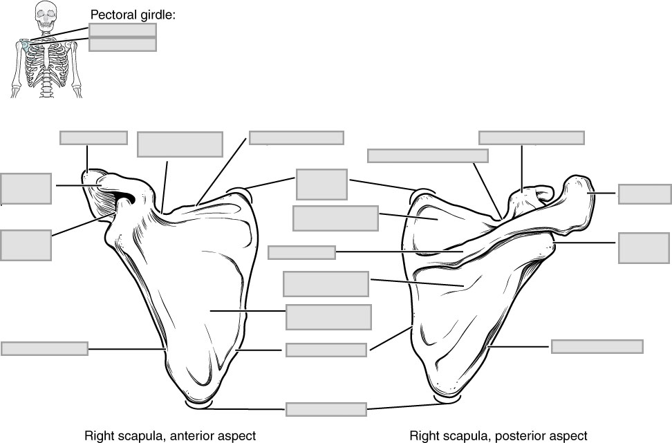
The scapula is also part of the pectoral girdle and thus plays an important role in anchoring the upper limb to the body (Figure 8.4). The scapula is located on the posterior side of the shoulder. It is surrounded by muscles on both its anterior (deep) and posterior (superficial) sides, and thus does not articulate with the ribs of the thoracic cage. Scapula forms the shoulder joint or glenohumeral joint involving articulation between the glenoid cavity of the scapula and the head of the humerus.
The Upper Limb
Each upper limb contains 30 bones. This includes the humerus, radius, ulna, carpals, metacarpals and the phalanges.
The Humerus
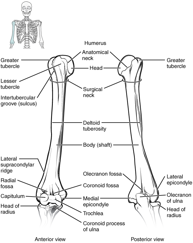
The upper limb is divided into three regions. These consist of the arm, located between the shoulder and elbow joints; the forearm, which is between the elbow and wrist joints; and the hand, which is located distal to the wrist. The humerus is the single bone of the upper arm region (Figure 8.5). At its proximal end is the head of the humerus. This is the large, round, smooth region that faces medially. The head articulates with the glenoid cavity of the scapula to form the glenohumeral (shoulder) joint.
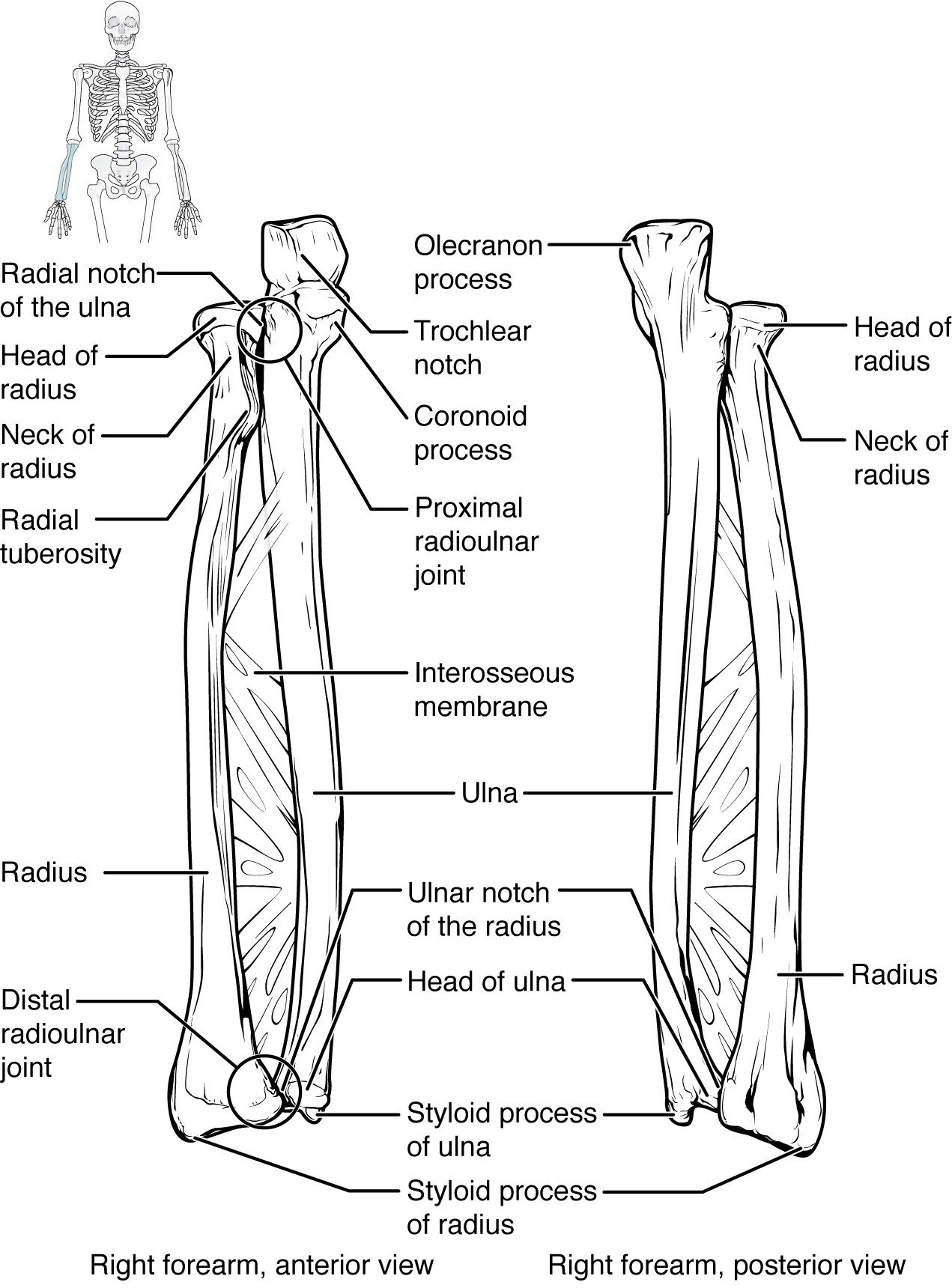
The Ulna and Radius
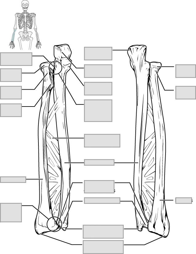
The ulna is the medial bone of the forearm. It runs parallel to the radius, which is the lateral bone of the forearm (Figure 8.6). The proximal end of the ulna resembles a crescent wrench with its large, C-shaped trochlear notch. This region articulates with the trochlea of the humerus as part of the elbow joint. The inferior margin of the trochlear notch is formed by a prominent lip of bone called the coronoid process of the ulna. The posterior and superior portions of the proximal ulna make up the olecranon process, which forms the bony tip of the elbow.
The radius runs parallel to the ulna, on the lateral (thumb) side of the forearm (see Figure 8.6). The head of the radius is a disc-shaped structure that forms the proximal end. The neck of the radius is the narrowed region immediately below the expanded head. The shaft of the radius is slightly curved and has a small ridge along its medial side. The distal end of the radius has a smooth surface for articulation with two carpal bones to form the radiocarpal joint or wrist joint.
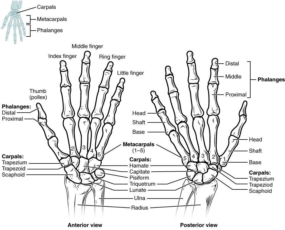
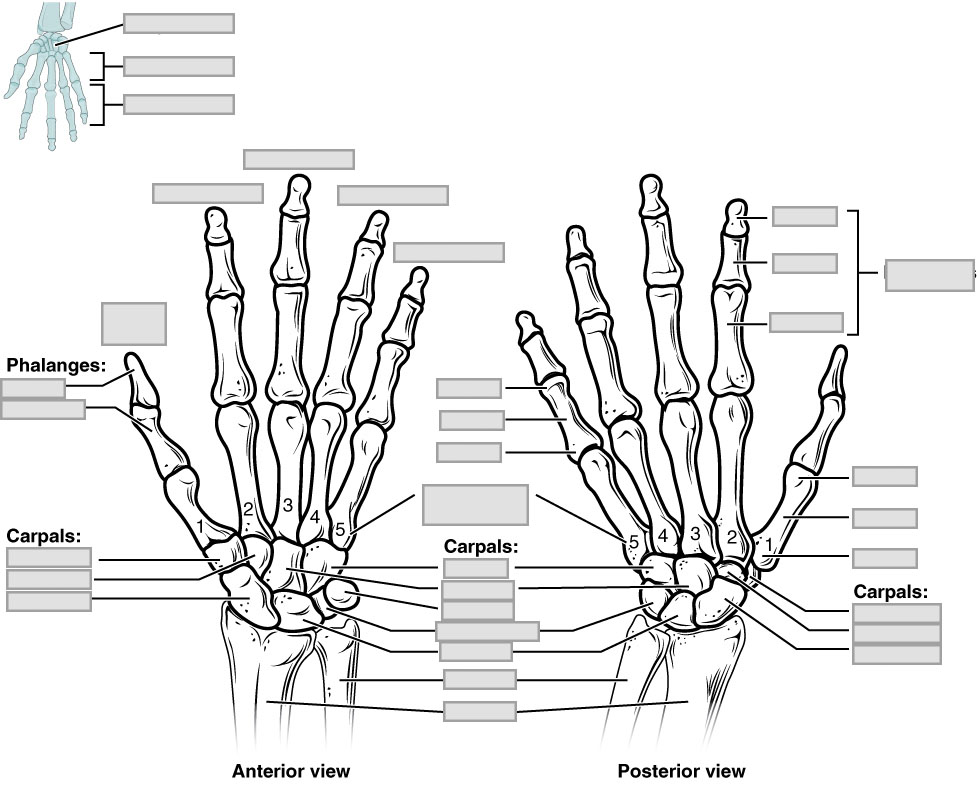
Bones of the Wrist and Hand
The wrist and base of the hand are formed by a series of eight small carpal bones (Figure 8.7). The carpal bones are arranged in two rows, forming a proximal row of four carpal bones and a distal row of four carpal bones. The bones in the proximal row, running from the lateral (thumb) side to the medial side, are the scaphoid, lunate, triquetrum and pisiform bones. The distal bones (lateral to medial) are the trapezium, trapezoid, capitate and hamate bones.
The palm of the hand contains five elongated metacarpal bones. These bones lie between the carpal bones of the wrist and the bones of the fingers and thumb (see Figure 8.7). The proximal end of each metacarpal bone articulates with one of the distal carpal bones.
The fingers and thumb contain 14 bones, each of which is called a phalanx bone, named after the ancient Greek phalanx. The thumb (pollex) is digit number 1 and has two phalanges, a proximal phalanx, and a distal phalanx bone (see Figure 8.7). Digits 2 (index finger) through 5 (little finger) have three phalanges each, called the proximal, middle, and distal phalanx bones.
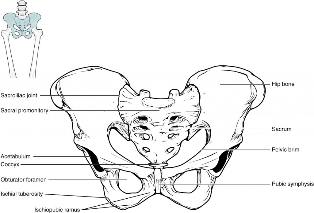
Bones of the Pelvic Girdle
The pelvic girdle (hip girdle) is formed by a single bone, the hip bone or coxal bone, which serves as the attachment point for each lower limb. Each hip bone, in turn, is firmly joined to the axial skeleton via its attachment to the sacrum of the vertebral column. The right and left hip bones also converge anteriorly to attach to each other (Figure 8.8).
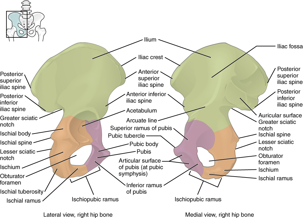
Os Coxa
The hip bone, or coxal bone, forms the pelvic girdle portion of the pelvis. The paired hip bones are the large, curved bones that form the lateral and anterior aspects of the pelvis. Each adult hip bone is formed by three separate bones that fuse together during the late teenage years. These bony components are the ilium, ischium, and pubis (Figure 8.9).
The ilium is the fan-like, superior region that forms the largest part of the hip bone. It is firmly united to the sacrum at the largely immobile sacroiliac joint (see Figure 8.8). The ischium forms the posteroinferior region of each hip bone. It supports the body when sitting. The pubis forms the anterior portion of the hip bone. The pubis curves medially, where it joins to the pubis of the opposite hip bone at a specialized joint called the pubic symphysis.
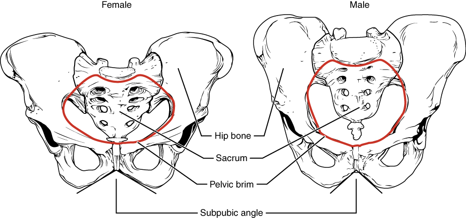
Male and Female Pelvis
The differences between the adult female and male pelvis relate to function and body size. In general, the bones of the male pelvis are thicker and heavier, adapted for support of the male’s heavier physical build and stronger muscles. The greater sciatic notch of the male hip bone is narrower and deeper than the broader notch of females. Because the female pelvis is adapted for childbirth, it is wider than the male pelvis, as evidenced by the distance between the anterior superior iliac spines (Figure 8.10). The ischial tuberosities of females are also farther apart, which increases the size of the pelvic outlet. Because of this increased pelvic width, the subpubic angle is larger in females (greater than 80 degrees) than it is in males (less than 70 degrees). The female sacrum is wider, shorter, and less curved, and the sacral promontory projects less into the pelvic cavity, thus giving the female pelvic inlet (pelvic brim) a more rounded or oval shape compared to males. The lesser pelvic cavity of females is also wider and more shallow than the narrower, deeper, and tapering lesser pelvis of males. Because of the obvious differences between female and male hip bones, this is the one bone of the body that allows for the most accurate sex determination.
The Lower Limb
Each lower limb contains 30 bones. This includes the femur, patella, tibia, fibula, tarsals, metatarsals and the phalanges.
The Lower Limb
The Femur
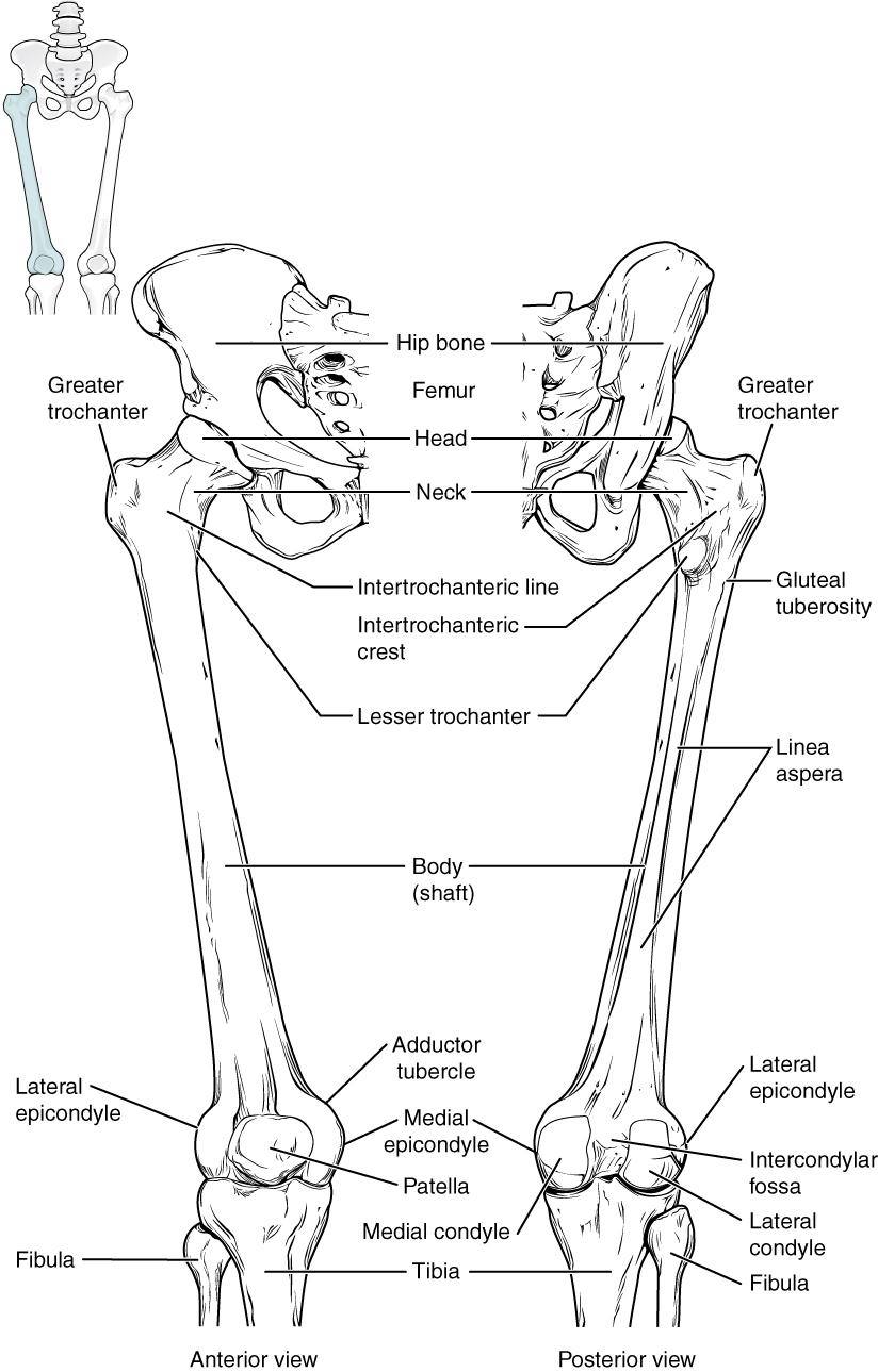
The femur, or thigh bone, is the single bone of the thigh region (Figure 8.11). It is the longest and strongest bone of the body, and accounts for approximately one-quarter of a person’s total height. The rounded, proximal end is the head of the femur, which articulates with the acetabulum of the hip bone to form the hip joint. The narrowed region below the head is the neck of the femur. The elongated shaft of the femur has a slight anterior bowing or curvature. The distal end of the femur has medial and lateral bony expansions. On the lateral side, the smooth portion that covers the distal and posterior aspects of the lateral expansion is the lateral condyle of the femur. The roughened area on the outer, lateral side of the condyle is the lateral epicondyle of the femur. Similarly, the smooth region of the distal and posterior medial femur is the medial condyle of the femur, and the irregular outer, medial side of this is the medial epicondyle of the femur. The combination of the medial and lateral condyles with the patellar surface gives the distal end of the femur a horseshoe (U) shape.
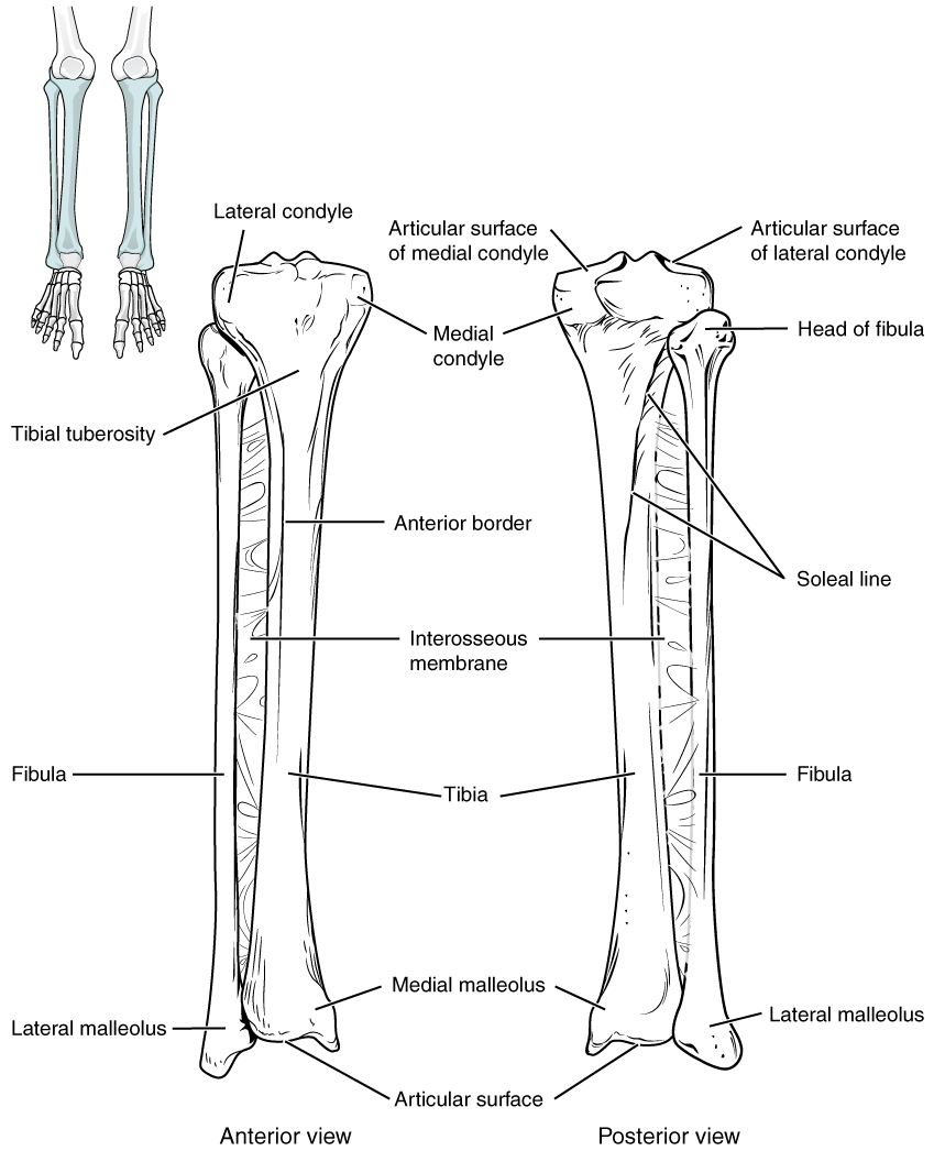
The Tibia and Fibula
The tibia (shin bone) is the medial bone of the leg and is larger than the fibula, with which it is paired (Figure 8.12). The tibia is the main weight-bearing bone of the lower leg and the second longest bone of the body, after the femur. The proximal end of the tibia is greatly expanded. The two sides of this expansion form the medial condyle of the tibia and the lateral condyle of the tibia.
The fibula is the slender bone located on the lateral side of the leg (see Figure 8.12). It serves primarily for muscle attachments and thus is largely surrounded by muscles. The head of the fibula is the small, knob-like, proximal end of the fibula. The distal end of the fibula forms the lateral malleolus, which forms the easily palpated bony bump on the lateral side of the ankle.
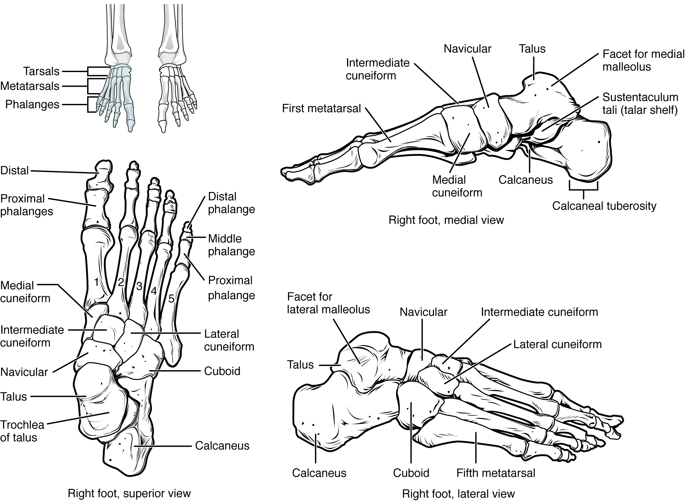
Bones of the Ankle and Foot
The posterior half of the foot is formed by seven tarsal bones (Figure 8.13). The most superior bone is the talus. This has a relatively square-shaped, upper surface that articulates with the tibia and fibula to form the ankle joint. fibula. Inferiorly, the talus articulates with the calcaneus (heel bone), the largest bone of the foot, which forms the heel.
The anterior half of the foot is formed by the five metatarsal bones, which are located between the tarsal bones of the posterior foot and the phalanges of the toes (see Figure 8.13). These elongated bones are numbered 1–5, starting with the medial side of the foot. The first metatarsal bone is shorter and thicker than the others. The second metatarsal is the longest.
The toes contain a total of 14 phalanx bones (phalanges), arranged in a similar manner as the phalanges of the fingers (see Figure 8.13). The toes are numbered 1–5, starting with the big toe (hallux). The big toe has two phalanx bones, the proximal and distal phalanges. The remaining toes all have proximal, middle, and distal phalanges.
Pre-Laboratory Questions
After reading the background information please answer these pre-laboratory questions.
- Name the main bones of the appendicular skeleton.
- What are the main bones in the pectoral girdle?
- Name the bones of the pelvic girdle.
- Name the long bones in the upper and lower limbs.
- What are the main wrist and finger bones called?
- What are the main ankle, foot and toe bones called?
Exercises
- Exercise 1 The Clavicle
- Exercise 2 The Scapula
- Exercise 3 The Upper Limb – Humerus
- Exercise 4 The Upper Limb – Ulna and Radius
- Exercise 5 Wrist and Hand
- Exercise 6 The Hip Bone (Ox Coxa)
- Exercise 7 Male and Female Pelvis
- Exercise 8 The Lower Limb – Femur
- Exercise 9 The Lower Limb – Tibia and Fibula
- Exercise 10 Ankle and Foot
Exercise 1 The Clavicle
Required Materials
- Clavicle from the disarticulated bones of the skeleton model
- Poster of the skeletal system
Procedure
- Obtain a clavicle and observe its structures from all sides (Figure 8.3)
- Identify the structures on the list below and label these on the figure given.
· Acromial end · Shaft · Coronoid tubercle · Sternal end · Costal tuberosity
Exercise 2 The Scapula
Required Materials
- Scapula from the disarticulated bones of the skeleton model
- Poster of the skeletal system
Procedure
- Obtain a scapula and observe its structures from all sides (Figure 8.4)
- Identify the structures on the list below and label these on the figure given.
· Acromion · Spine · Glenoid cavity · Subscapular space · Infraglenoid tubercle · Supraglenoid tubercle · Infraspinous process · Superior angle · Inferior angle · Superior border · Lateral border · Suprascapular notch · Medial border · Supraspinous fossa
Exercise 3 The Upper Limb – Humerus
Required Materials
- Humerus from the disarticulated bones of the skeleton model
- Poster of the skeletal system
Procedure
- Obtain a humerus and observe its structures from all sides (Figure 8.5)
- Identify the structures on the list below and label these on the figure given.
· Anatomical neck · Lesser tubercle · Capitulum · Lateral supracondylar ridge · Coronoid fossa · Medial epicondyle · Deltoid tuberosity · Olecranon fossa · Greater tubercle · Radial fossa · Head · Shaft · Intertubercular groove · Supracondylar ridges · Lateral epicondyle · Surgical neck
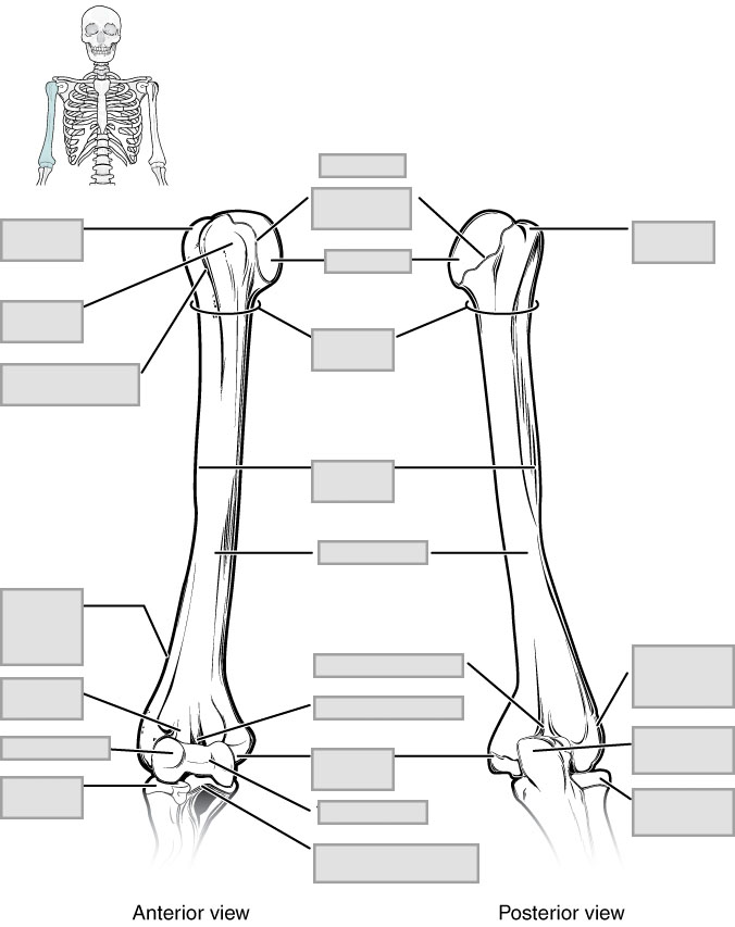
Exercise 4 The Upper Limb – Ulna and Radius
Required Materials
- Radius from the disarticulated bones of the skeleton model
- Ulna from the disarticulated bones of the skeleton model
- Poster of the skeletal system
Procedure
- Obtain a radius and an ulna and observe its structures from all sides (Figure 8.6)
- Identify the structures on the list below and label these on the figure given.
· Coronoid process · Radial tuberosity · Head of radius · Radius · Head of ulna · Styloid process of radius · Neck of radius · Styloid process of ulna · Olecranon process · Trochlear notch · Radial notch of ulna · Ulna
Exercise 5 Wrist and Hand
Required Materials
- Wrist and hand from the disarticulated bones of the skeleton model
- Wrist and hand on the articulated skeleton model
- Poster of the skeletal system
Procedure
- Examine the bones of the wrist and hand from the whole skeleton or a model and observe their structures from all sides (Figure 8.7)
- Identify the structures on the list below and label these on the figure given.
· Carpals · Metacarpal IV · Distal phalanx · Metacarpal V · Distal phalanx (pollex) · Middle phalanx · Metacarpal I · Proximal phalanx · Metacarpal II · Proximal phalanx (pollex) · Metacarpal III
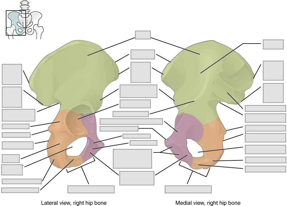
Exercise 6 The Hip Bone (Ox Coxa)
Required Materials
- Hip bone from the disarticulated bones of the skeleton model
- Hip bone on the articulated skeleton model
- Poster of the skeletal system
Procedure
- Examine the hip bone from the whole skeleton or a model and observe their structures from all sides (Figure 8.9)
- Identify the structures on the list below and label these on the figure given.
· Acetabulum · Ischial sciatic notch · Anterior inferior iliac spine · Ischial spine · Anterior superior iliac spine · Ischial tuberosity · Arcuate line · Ischium · Arcuate surface · Lesser sciatic notch · Greater sciatic notch · Obturator foramen · Iliac crest · Posterior inferior iliac spine · Iliac fossa · Posterior superior iliac spine · Ilium · Pubic body · Ischial body · Pubis
Exercise 7 Male and Female Pelvis
Required Materials
- None
Procedure
- Examine both the male and female pelvises and observe their structures from all sides (Figure 8.10)
- Identify the structures in the list below and label these on the figure given.
· Hip bone · Pelvic inlet · Obturator foramen · Sacrum · Pelvic brim · Subpubic angle 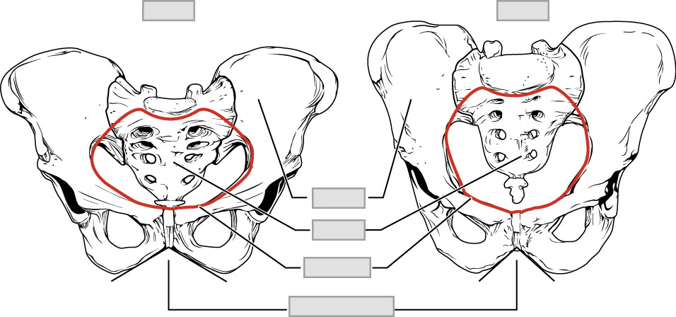
Exercise 8 The Lower Limb – Femur
Required Materials
- Femur from disarticulated skeleton model
- Femur on articulated skeleton model
- Poster of the skeletal system
Procedure
- Examine an isolated femur and observe its parts from all sides (Figure 8.11)
- Identify the structures in the list below and label these on the figure given.
· Body (shaft) · Lateral epicondyle · Gluteal tuberosity · Medial condyle · Greater trochanter · Medial epicondyle · Head · Neck · Lateral condyle

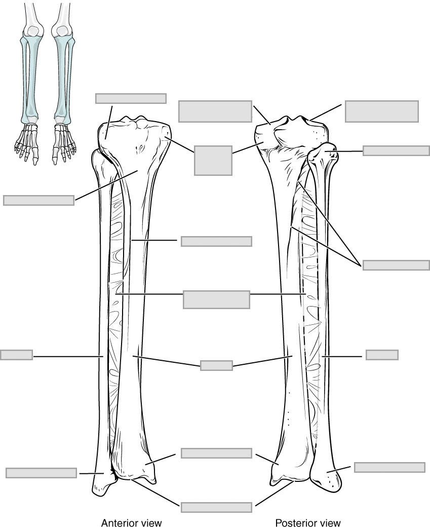
Exercise 9 The Lower Limb – Tibia and Fibula
Required Materials
- Tibia and fibula from disarticulated skeleton model
- Tibia and fibula on articulated skeleton model
- Poster of the skeletal system
Procedure
- Examine an isolated tibia and a fibula and observe its parts from all sides (Figure 8.12)
- Identify the structures in the list below and label these on the figure given.
· Articular surface · Lateral malleolus · Articular surface of lateral epicondyle · Medial condyle · Fibula · Medial malleolus · Head of fibula · Tibia · Interosseous membrane · Tibial tuberosity · Lateral condyle
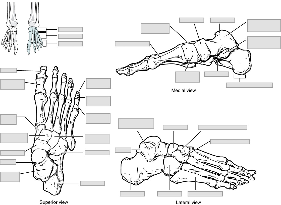
Exercise 10 Ankle and Foot
Required Materials
- Foot and ankle bones from disarticulated skeleton model
- Foot and ankle on articulated skeleton model
- Poster of the skeletal system
Procedure
- Examine the foot and ankle bones from the models and observe its parts from all sides (Figure 8.13)
- Identify the structures in the list below and label these on the figure given.
Calcaneus Metatarsal 4 Distal phalanges Metatarsal 5 Metatarsal 1 Middle phalanges Metatarsal 2 Proximal phalanges Metatarsal 3 Talus
Post-laboratory Questions
- What are some similar features of the upper limb and lower limb long bones?
- How are the two main bone types of the pectoral girdle (clavicle and scapula) different than the main bones of the pelvic girdle?
- Compare the organization of the hand bones and foot bones. What are some similarities and differences?
