
21 Chapter 21 The Lymphatic and Immune System
By Joseph D’Silva
Motivation.
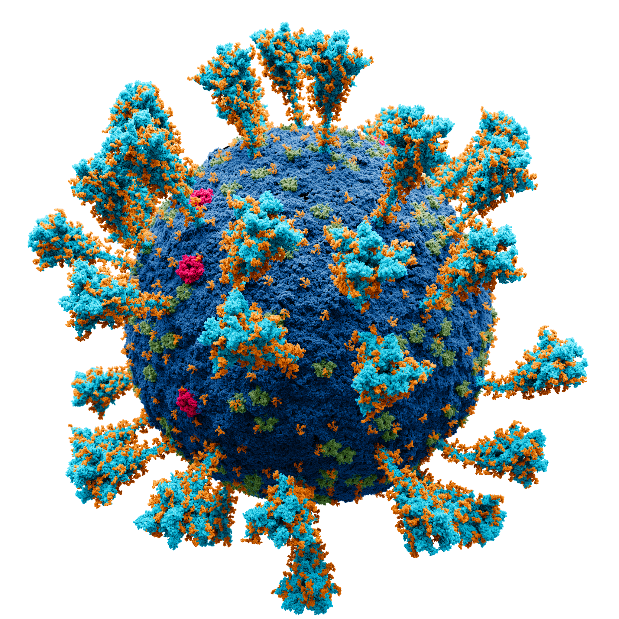
Vaccination has been very much in the news since the novel coronavirus SARS-CoV-2 messed up our lives beginning in December 2019. There was a race to create and manufacture a vaccine against the virus quickly because thousands of lives were being lost daily worldwide. Finally, in 2020, vaccines were developed and have been a savior for people. In 2022, people above 55 years of age are able to get their fourth vaccine shot for the Corona virus.
Vaccination is related to immunity. It provides protection to the body so that an infectious agent does not attack it. Every year, people get vaccinated against the flu (influenza) virus. Vaccinations protect us also against polio, mumps, smallpox and other diseases. Immunity is a part of the lymphatic system in our body. To understand the lymphatic system, we will have to know its structure and how it functions physiologically.
Learning Objectives
Upon completion of the work in this chapter students should be able to:
- Identify and describe the structure and distribution of major lymphatic vessels and organs
- Describe the histology of a lymph node, spleen, tonsils, Peyer’s patches
- Compare and contrast the structure and function of different lymphoid organs
Background.
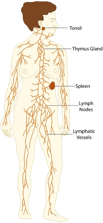
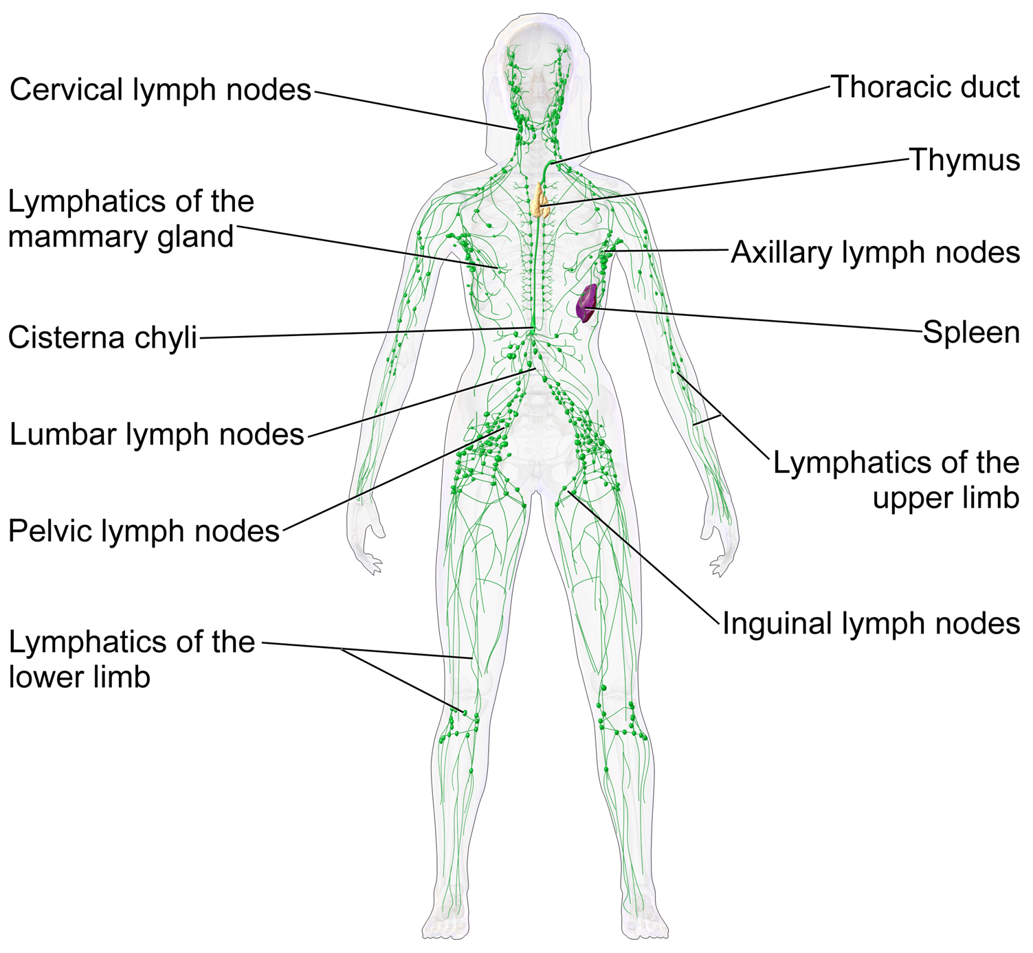
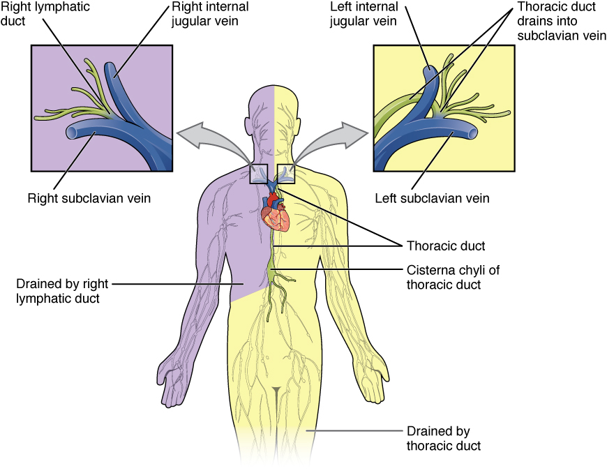
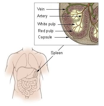
The lymphatic system is made up of lymph fluid, cells, tissues and organs. Lymph is a colorless fluid that contains cells. The cells are lymphocytes. They are found in lymph nodules and nodes and in spleen, bone marrow and thymus (Figure 21.2 and 21.3). Lymphocytes proliferate in the tissues and mature there. Lymphatic vessels (capillaries) are structures through which cells travel and can be found under the skin. These capillaries have closed or blinded ends. Vessels are organized into larger trunks. The trunks are (1) right and left jugular; (2) right and left subclavian; (3) right and left bronchomediastinal; (4) intestinal; (5) right and left lumbar trunks. Each trunk drains a particular area in the body. The trunks lead to two ducts: (1) right lymphatic and (2) thoracic (Figure 21.4).
White blood cells travel through the lymphatic ducts going to tissues and organs where they are needed to neutralize harmful pathogens (antigens). These cells are macrophages, B-lymphocytes and T-lymphocytes. When an antigen is detected, B-lymphocytes react to it and produce antibodies. The antibodies provide us with immunity and are produced by the T-lymphocytes and B-lymphocytes after a person receives vaccinations. Vaccines used in vaccination are prepared from dead infectious agents, for example, COVID virus. Lymphatic tissue is found in these organs: thymus, spleen and ileum.
Histology of Lymphatic Organs
Thymus (Figure 21.5). The thymus consists of two lobes, merged in the middle, surrounded by a capsule. The lobes consist of an outer cortex rich with cells and an inner less dense medulla. The lobes are divided into smaller lobules between which trabeculae radiate out from the capsule along the division lines of the lobes.
The cortex is mainly made up of thymocytes and epithelial cells. The thymocytes, immature T cells, are supported by a network of the finely-branched epithelial reticular cells, which is continuous with a similar network in the medulla. This network forms an adventitia to the blood vessels, which enter the cortex via divisions (septa) near the junction with the medulla. Other cells are also present in the thymus, including macrophages, dendritic cells, and a small amount of B cells, neutrophils and eosinophils.
In the medulla, the network of epithelial cells is coarser than in the cortex, and the lymphoid cells are relatively fewer in number. Concentric, nest-like bodies called Hassall’s corpuscles (also called thymic corpuscles) are formed by aggregations of the medullary epithelial cells. These are concentric, layered whorls of epithelial cells that increase in number throughout life.
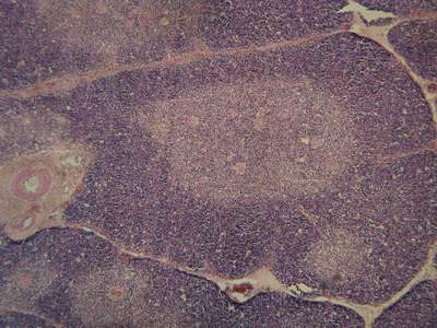
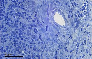
Spleen (Figure 21.6) The spleen contains two different tissues, white pulp and red pulp. The white pulp functions in producing and growing immune and blood cells. The red pulp functions in filtering blood of antigens, microorganisms, and defective or worn-out red blood cells. The white pulp contains many lymphocytes which are small cells with dark purple nuclei in H&E stained slides. Most of the rest of the mesenchyme of the spleen is red pulp. You can also observe the dividing connective tissue or trabeculae that extend in from the capsule covering of the spleen.
Tonsils (Figure 21.7) Tonsils are large non-encapsulated (or partially encapsulated) masses of lymphoid tissue. They can be found in the pharynx or nasopharynx and at the base of the tongue. The superficial part of the tonsils are covered with a stratified squamous epithelium. Tonsils have many invaginations which form blind crypts. Below the epithelium, there are many lymphoid follicles beneath which have germinal centers.
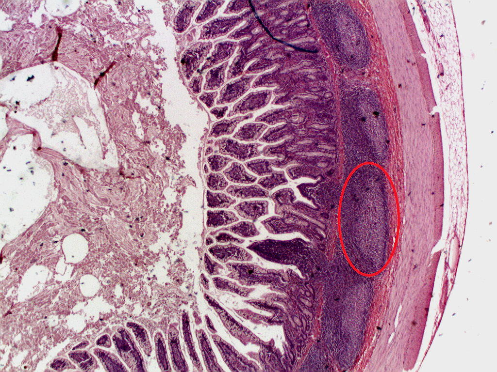
Peyer’s patches (Figure 21.8) Peyer’s patches, which are organized lymphoid nodules, are commonly found in the small intestines. Peyer’s patches appear as oval or round lymphoid follicles extending from the mucosa layer of the ileum into the submucosa layer. B lymphocytes found in the follicles’ germinal centers and these lymphocytes are the majority in adults. T lymphocytes are found in the zones between follicles. Follicle-associated epithelium covers all lymphoid follicles which differ from the intestinal epithelium of the villi due to having fewer goblet cells. This layer contains M cells or microfold cells which uptake and transport of antigens from lumen.
Pre-Laboratory Questions
After studying the Background information above answer the following questions prior to doing the lab exercises.
1. Define lymphocytes, lymph and lymphatic capillaries.
2. Sketch a figure to show the lymphatic trunks and ducts.
3. List the areas of the body that are drained by each individual trunk.
4. Describe the structures in a spleen section that you would find under the microscope.
5. Compare and contrast the histological structure of the thymus and a tonsil.
Exercises
- Exercise 1 Identify the main organs and glands of the lymphatic system
- Exercise 2 Histology of the thymus
- Exercise 3 Microanatomy of the spleen
- Exercise 4 Examination of tonsil histology
- Exercise 5 Vermiform appendix / ileum Peyer’s patches
Exercise 1 Identify the main organs and glands of the lymphatic system
Required Materials
- Torso models
- Lymphatic System Anatomy poster
- The Body’s Defenses poster
- Post-it notes
- Labeling tape
Procedure
- Use your textbook, torso models, lymphatic system anatomy poster, and the body’s defenses poster to identify the structures of the immune system shown in Figure 21. 2, 21.3 and 21.4.
- Locate the thymus gland and spleen. The thymus gland can be found in the mediastinum. The spleen is located in the ULC (upper left quadrant) in the pelvic cavity. The vermiform appendix is in the RLQ (right lower quadrant). All these organs contain lymphatic tissue.
- Use post-it notes or labeling tape to label as many of these lymphoid tissues, organs, and vessels as you can find on the torso models.
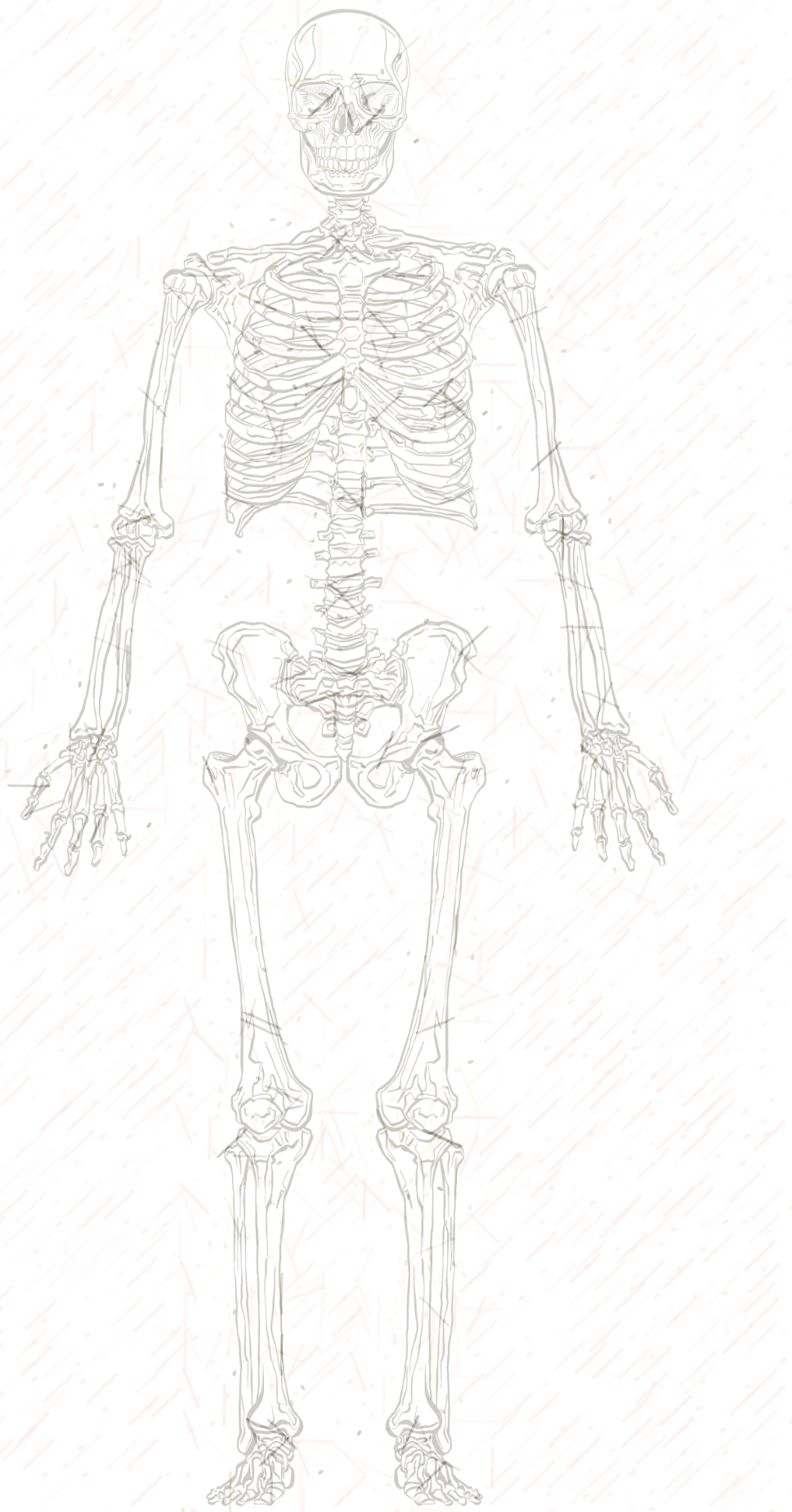
- Take a picture of the torso model with the post-it notes/labels. Insert your picture in the space below. Alternatively, you can sketch the body (use the skeletal outline given as a guide), and label these lymphoid structures.

Exercise 2 Histology of the thymus
Required Materials
- Compound microscope
- Microscope lens paper
- Microscope lens cleaning solution
- Microscope immersion oil
- Slide of the Human Thymus
Procedure
- Ask your instructor for a slide showing the thymus (Figure 21.5).
- Scan under low power.
- The tissue in the thymus is divided into lobules. The lobules are separated from one another by trabeculae. Identify trabeculae which appear as thin white connective tissue. Each lobule consists of a light central medulla and a cortex that surrounds it. The cortex consists of a dense population of T-lymphocytes in stages of development and appears blue in color.
- Select different regions of the thymus and observe under high power.
- Take a picture of your observations at low power and high power, paste the picture below and label the main structures and cell types observed.
Exercise 3 Microanatomy of the spleen
Required Materials
- Compound microscope
- Microscope lens paper
- Microscope lens cleaning solution
- Microscope immersion oil
- Slide of the Human Spleen
Procedure
- Ask your instructor for a slide showing the spleen (Figure 21.6).
- Scan under low power.
- The tissue in the spleen can be identified by its white and red pulp. The white pulp surrounds an artery and consists of B-lymphocytes and T-lymphocytes mostly. The red pulp is made up of red blood cells. Trabeculae can be observed in the the splenic tissue.
- Select different regions of the spleen and observe under high power.
- Take a picture of your observations at low power and high power, paste the picture below and label the main structures and cell types observed.
Exercise 4 Examination of tonsil histology
Required Materials
- Compound microscope
- Microscope lens paper
- Microscope lens cleaning solution
- Microscope immersion oil
- Slide of the Human Palatine Tonsil
Procedure
- Ask your instructor for a slide showing a tonsil (Figure 21.7).
- Scan under low power.
- Tonsils are lymphatic nodules. They are located in the oral cavity and pharynx. The tissue is broken up by a number of crypts. These are deep infolding tissue. Tonsilar nodules are made up of large number of lymphocytes. Germinal centers are light in color and surrounded by lymphocytes that are deep blue.
- Select different regions of the tonsil and observe under high power.
- Take a picture of your observations at low power and high power, paste the picture below and label the main structures and cell types observed.
Exercise 5 Vermiform appendix / ileum Peyer’s patches
Required Materials
- Compound microscope
- Microscope lens paper
- Microscope lens cleaning solution
- Microscope immersion oil
- Slide of the Human Ileum with Peyer’s Patches
- Ask your instructor for a slide showing the vermiform appendix/ileum (Figure 21.8).
- Scan under low power.
- Located in the submucosa are a number of lymphatic nodules. Each nodule consists of a light germinal center surrounded by darker staining capsule. Peyer’s patches are nodules in the walls of the ileum. They are made up of lymphocytes.
- Select different regions of the Peyer’s patches and observe under high power.
- Take a picture of your observations at low power and high power, paste the picture below and label the main structures and cell types observed.
Post-laboratory Questions
- The areas of the body that are drained by the right and left subclavian trunks are the _____limbs.
- Two lymphatic ducts are the ________ and _______.
- Sketch the structure of spleen and describe it.
- Study the structure of a tonsil and describe it.
- What are Peyer’s patches and where are they found?
