
4 Chapter 4 The Tissue Level of Organization
By Joseph D’Silva
Motivation
Diabetes is a disorder of the endocrine system. It is caused by a more-than-normal level of glucose sugar in the blood. The normal level is 70-100 mg/dL. Above 100 mg/dL, the person has an impaired blood-glucose level. There is a high incidence of diabetes among Afro-Americans. 34.2 million people of all ages—or 10.5% of the US population—has diabetes. Among African Americans, 10.9 % are affected. About 7.3 million adults aged 18 years or older are not aware of diabetes.
The amount of blood sugar in the body is regulated by insulin. This is a hormone and is secreted by cells known as the Islets of Langerhans that are located in the tissues of the pancreas. When the islets release insulin, blood sugar is controlled in the blood. When it fails to, a person develops diabetes.
To know about the pancreas is to study its histology. The histological examination of a pancreas requires that it be sectioned by using a microtome. (A microtome is an instrument. You can slice tissues as thin as 05-06 microns. A micron ((symbol u (mu)) is 1/000 mm. Look up mm lines on your ruler.) The slices of tissues can be processed through a procedure and stained with dyes. After staining and more processing they can be put on a glass slide with a cover slip on top and sealed with a sealant (Canada balsam or Permount). Once dried, the section is then examined under a compound microscope using 10 x 4, 10 x 10, or 10 x 40 magnification.
This laboratory exercise studies a section of the pancreas to identify structures in it. The structures are:
- Lobules
- Pancreatic acini
- Islets of Langerhans
See figures below and locate the (a) pancreas; (b) Islets of Langerhans; (c) Interlobular ducts, acinar cells.

Learning Objectives
Upon completion of work in this chapter, students should be able to:
- Define histology and histological tissues in general.
- Define and recognize four types of tissues on stained tissues on microscope slides.
- Describe tissues of organs.
- Explain a histologically impaired tissue.
Background
There are more than 200 different tissue types in the human body. All the organs in the body are made of tissues that are made of cells. The tissues in a body characterize organs. It would be impossible to try and remember all the tissues that are characteristic of all the organs. Therefore, histologists (cell-study specialists) have classified the 200 tissues into four categories: Epithelial, Connective, Muscle and Neural. Each category of tissues has its own distinct characteristics. For example, epithelial cells are flat, cuboidal or tall. Connective tissue can be tough or fluid. Muscle tissue is made of muscle fibers (cells). Nerve tissues is made up of neurons.
Consider your skin or integument. If you cut yourself or if a surgeon makes an incision on your skin you will see tissues that are epithelial, connective, muscular and neuronal. The first layer is the epithelial tissue made up of epithelial cells. The second layer is connective. There are blood vessels and fibrous tissues there. Interspersed in the connective tissue are nerve cells. Below this are fat cells. Finally, there is a layer of muscle tissue.
In this chapter, you will learn to recognize cell types and to identify tissues. You will need to work with a microscope and study prepared slides using 40x, 100x and 400x magnification.
Epithelial Tissues
Epithelial tissue appears in sheets of cells. It covers body surfaces, lining of internal surfaces and forms glands. Cells that make up the epithelial tissue have an apical and basal surface (aka polarity), and lateral surfaces that include tight junctions. The basal layer is attached to basement membrane (connective tissue) beneath it. Another characteristic of epithelial tissue is avascularity, meaning there is no blood supply to it. Epithelial tissue is classified by (a) shape, and (b) layers.
a. Classification by shape:
Squamous – Cells are thin and flat; look like fried eggs with sunny side up or tiles on a floor or fish scales; imagine the yolk to be the nucleus and the white to be cytoplasm of epithelial cells. The nucleus is central, disc-like with cytoplasm around it. Examples are: air sacs of lungs (Figure 4.2) , blood vessels (arteries and veina), body cavity and lymphatic vessels.

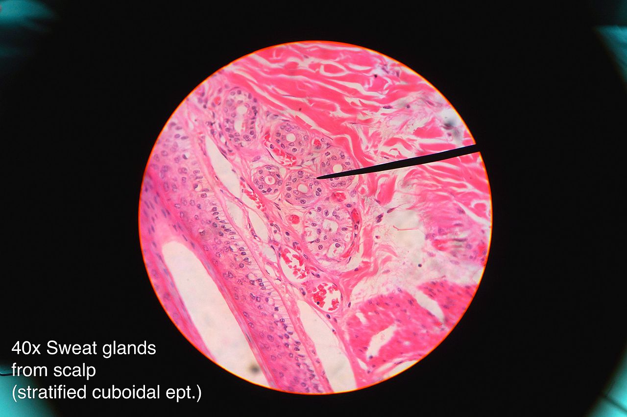
Cuboidal – Cells are shaped like cubes with the length, breadth and height equal in size. The nucleus is situated in the cell center with cytoplasm equally distributed right round. Examples: kidney tubules, ducts of small glands (Figure 4.3), ovary surface.
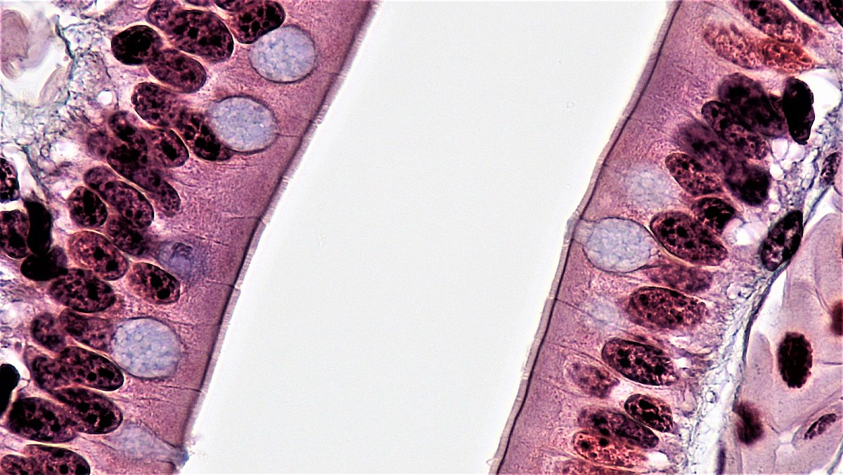
Columnar – Cells are shaped like tall columns. The height is longer than width. The nucleus is round or oval and is situated closer to basal surface. Nuclei may be oval/round nuclei and appear in a row. This is a characteristic feature of columnar epithelium. Examples: intestine (cells are interspersed with epithelial goblet cells) (Figure 4.4), gallbladder, uterus.
b. Classification by cell layers
Simple epithelium is one cell thick layer and is in direct contact with basement membrane. Its functions are absorption; diffusion, filtration or secretion. Figures 4.2, 4.3, and 4.4 all show simple epithelia.
Stratified Epithelium is two or more layers of cells thick. The deepest layer makes contact with basement membrane. Its functions are: protection/resist abrasion.
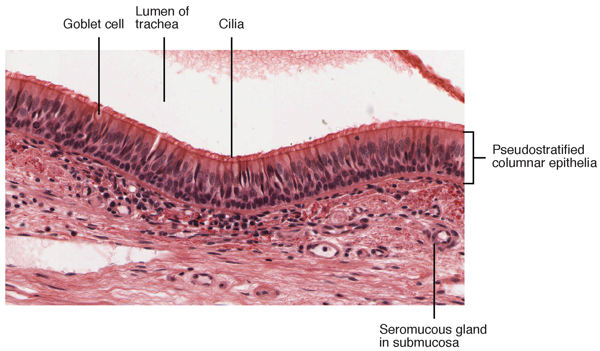
Pseudostratified Epithelium. The tissue appears in strata because cell nuclei are situated at a distance from the basal surface. But all the epithelial cells make direct contact with basement membrane. Its functions are absorption or secretion. Examples are trachea (Figure 4.5), epididymis.
Connective Tissues
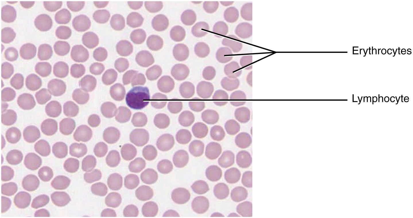
Connective tissue is totally different from epithelial tissue. Connective tissue consists of cells plus extracellular matrix. The structure in which the cells are embedded is the extracellular matrix. For example, blood cells are found in matrix called plasma (fluid). Extracellular matrix contains solid collagen and reticular fibers for many cells. In connective tissues, the matrix is the predominant structure with cells being sparse. The matrix consists of ground substance and protein fibers. The ground substance and fibers are secreted by the sparse cells. Blood (fluid), cartilage (semi-solid) and bone (solid) are connective tissues. The function of connective tissue is to connect tissues. For example, blood is pumped from the heart to all parts of the body and connects one tissue with another.
Connective Tissue Cells: Associated with connective tissue are several types of cells called fibroblasts, macrophages, adipocytes, mast cells and stem cells. They are known as resident cells because they are permanent where they be found. On the other hand, lymphocytes, plasma cells, neutrophils, eosinophils, basophils and monocytes are called wandering cells because they can move. Each cell is different from the other and has specific functions.
Classification
Connective tissue is classified as (a) Embryonic Connective Tissue; (b) Connective Tissue Proper; and, (c) Specialized Connective Tissue.
Embryonic Connective Tissue is derived from the embryo. Mesenchyme is connective tissue that is found in the embryo. Mesenchymal CT: Mesenchymal cells, ground substance and fibers make up embryonic connective tissue. The cells are few; ground substance is fluid-like and the fibers are few. Cells are spindle shaped. Mucous connective tissue is present in the umbilical cord. Wharton’s jelly is the ground substance in which spindle-shaped cells are found.
Connective Tissue Proper is different from Specialized Connective Tissue. It is divided into Loose connective tissue and Dense connective tissue. Here, you will have to identify three types of fibers: collagen , elastic and reticular fibers. Collagen and reticular fibers are made up fibrous collagen proteins whereas elastic fiber consists of fibrous elastin protein. The three types of fibers give tensile strength to tissues. Connective tissue proper contains much fibers compared to Specialized Connective Tissue.
Loose Connective Tissue does not have much collagen fibers. Ground substance occupies much of the space in the tissue. There are few transient, wandering cells. Loose connective tissue can also be described as (a) Areolar tissue; (b) Adipose (fat) tissue; and, (c) Reticular tissue. Loose connective tissue is found underneath the skin and the lining of internal surfaces.
Dense Connective Tissue. As the name suggests, this tissue type is dense. It is pretty heavy with collagen fibers. Cells called fibroblasts are few. Fibroblast cells secrete the collagen fibers. Dense connective tissue is also found under the skin. Tendons, ligaments and aponeuroses are made up of dense connective tissue.
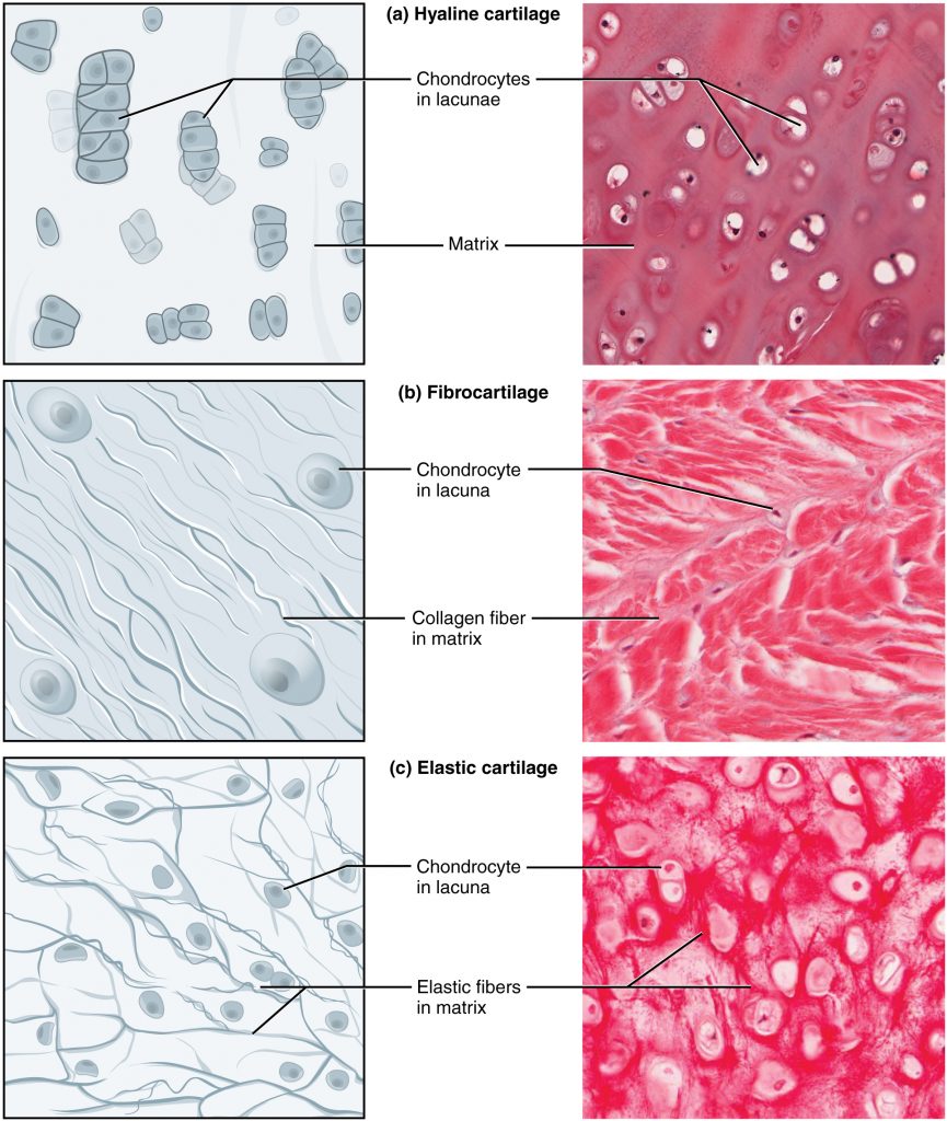
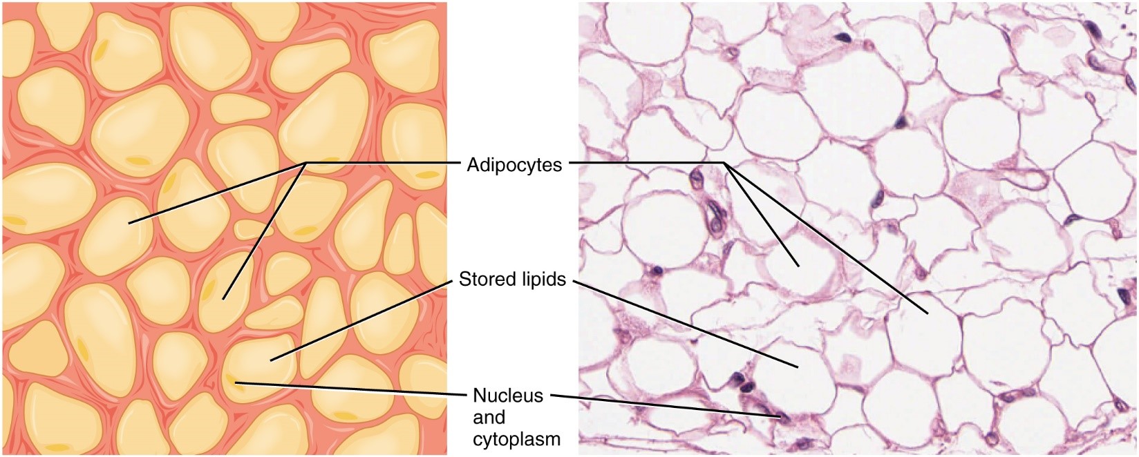
Specialized Connective Tissue. These connective tissues comprise cartilage, bone and fat tissue. Cartilage is made up of semi-solid structures (Figure 4.9) while bone is solid due to calcium salts. Fat tissue contains adipocyte cells filled with droplets of fat pushing the nucleus to the side (Figure 4.10).
Muscle Tissues
Muscle tissue is made up of muscle cells, also known as muscle fibers (Please remember!). Muscle cells are different from epithelial cells and connective tissue cells. They are long (because they have to stretch), have a nucleus and make up the bulk of a human body.
There are three types of muscle cells: skeletal, cardiac and smooth (Figure 4.11). Each type is remarkably different from the other.

Skeletal muscle fibers (cells) are long, have more than one nucleus (multinucleate) and striations (stripes) in the cytoplasm. Muscle cells bind bone to bone. Cardiac muscle fibers are branched, striated but with one nucleus per cell and have intercalated discs. Intercalated discs are unique to cardiac muscle and are junctions between the cells. Cardiac cells are found in heart tissue only. Smooth muscle fibers are pointed at two ends but with a bulge in the middle (spindle-shaped) with one nucleus per cell. They do not have striations or intercalated discs or branches. Smooth muscles are found in the wall of the stomach, intestine, sphincters and the eye.
Function: Skeletal muscles attach bone to bone. They move bones. Every movement in our skeletal system is made by skeletal muscles. It functions voluntary, that is, we can will our bones to move. Cardiac muscles and Smooth Muscles are involuntary; we have no control over them from the time we are born to the time we die.
Nervous Tissues
Nervous tissue is made up of large neural cells (neurons) and supporting cells (neuroglia). A neuron has one nucleus in its cytoplasm. The shape of nerve cells is irregular. The cytoplasm extends itself into branches. The ends of the branches come into contact with those of other nerve cells and pass on sensory stimuli. The branches that receive stimuli (such as heat, light, sound) are called dendrites. The stimuli pass through the cytoplasm and are relayed through branches known as axons. Axon ends synapse (form junctions) with dendrites, cell bodies, or other axons to pass on the stimuli to billions of nerve cells that are found in our brain and throughout the body. The stimuli are important to us because they allow the body to coordinate our physiological functions. Neuroglia (aka glial cells) are smaller cells that support and protect neurons.
Pre-Laboratory Questions
Review the background information sections on tissues that are provided in this chapter prior to attending the laboratory. Then, answer the following questions:
- Define tissues.
- List the four categories of tissues. For each category, give an example of an organ where you would find the tissue.
- What are types of shapes that define epithelial tissues by?
- Name epithelial tissues according to their layers. Cite one example for each.
- Compare and contrast stratified epithelium with pseudostratified epithelium.
Exercises
- Exercise 1. Epithelial Tissues
- Exercise 2. Connective Tissues
- Exercise 3. Muscle Tissues
- Exercise 4. Neural Tissues
- Exercise 5. Histologically Impaired Tissues
Exercise 1. Epithelial Tissues
Required Materials
- Compound microscope
- Lung tissue slide
- Slide of skin from general surface showing sweat glands
- Slide of columnar epithelium from small intestine
- Trachea slide
- Adipose tissue slide
- Muscle slide with skeletal, cardiac and smooth muscle
- Spinal cord slide
- Pancreas slide
Exercise 1.1. Identification of Squamous Epithelial Tissue.
This is what you should know and remember about squamous epithelial tissue: Cells look like fried eggs with nucleus like yolk sitting on top of cytoplasm (white of egg), sunny-side up, or tiles on a floor. Filtration (glomerulus), diffusion of gases (lung tissue).
This is what you should do:
- Ask your instructor for a prepared slide showing lung tissue. (Figure. 4.2.)
- Place it on the stage of microscope and bring it into focus using low power (4 x 10 magnification).
- Study tissue on slide. Move slide vertically and horizontally. Most of the tissue will appear as large, irregular white spaces encircled by thin layer of cells. The spaces are alveolar sacs. The cells are squamous epithelial.
- The squamous epithelial cells are tiny but with a prominent blue (or dark) nucleus. You will not be able to see the margins of the cells because they are indistinct.
- Change the magnification to high power (10 x 10). Study the prominent, blue or dark nuclei of the cells. Each nucleus is surrounded by cytoplasm. The plasma membrane is very thin. It will not appear as a distinct structure.
- Identify alveolar space and nucleus of the squamous epithelial cell. Use Fig. 1.2 to assist you.
- Draw the tissues at low and high magnification to show the cell and tissue structure as best as you can. Label nucleus, cytoplasm.
Exercise 1.2. Identification of Simple Cuboidal Epithelium
This is what you should do:
- Ask your instructor for a slide showing sweat gland tissue or kidney tubule. (Figure 4.3.)
- Place it on the stage of the microscope and bring the tissue into focus using low power (4 x 10) magnification.
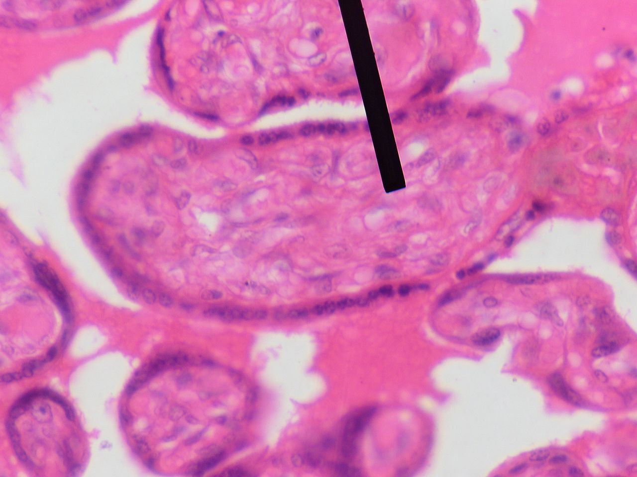
- Move slide horizontally and vertically to zoom in on cells (see black label). The space in the center of the tubule is the lumen. Cuboidal cells circle the lumen. The cells are more or less all the same shape with a large nucleus in the center. The sides of the cells are equal in length, breadth and height.
- The side of cell facing the lumen is the apical surface. The side away from it is the basal surface.
- Change magnification to high power (10 x 10) magnification. The cells will appear larger. The cytoplasm and nucleus now appears more distinctly.
- Draw what you observe at low and high magnification and label lumen, nucleus, cytoplasm, apical and basal surface.
Exercise 1.3. Identification of Simple Columnar Epithelium
This is what you should do:
- Ask your instructor for a slide showing small intestine tissue. (Figure 4.4.)
- Place it on the stage of the microscope and bring the tissue into focus using low power (4 x 10) magnification.
- Move slide horizontally and vertically to zoom in on a piece of the tissue that looks like a finger. The cells are numerous and colored light blue/purple with a prominent nucleus. They appear on the two sides of the intestinal tissue. The cells are taller than they are wide in shape with a large nucleus close to the base. In between the tall cells are goblet cells. They look like small vases and are filled with mucus.
- Change magnification to high power (10 x 10) magnification. The cells will appear larger. The cytoplasm and nucleus now appears more distinctly. The space between the two sides with cells contains blood vessels and nerve fibers.
- Draw what you observe at low and high magnification, and label lumen, nucleus, cytoplasm, apical and basal surface.
Exercise 1.4. Pseudostratified Columnar Epithelium
This is what you should do:
- Ask your instructor for a slide showing trachea. (Figure 4.5.)
- Place it on the stage of the microscope and bring the tissue into focus using low power (4 x 10) magnification.
- Move slide horizontally and vertically to zoom in on cells that appear in two or more layers. Cells are tall but blunt towards apical surface. Not all cells reach up to surface and are shorter, situated halfway between the taller ones. This gives a false impression there are two layers of cells. That is why they are called pseudostratified. The cells are numerous and colored light blue/purple with a prominent nuclei. The nuclei are prominent.
- Change magnification to high power (10 x 10) magnification. Notice that all the cells — tall or short — all share same basement membrane.
- Draw what you observe at low and high magnification, and label lumen, nucleus, cytoplasm, apical and basal surface.
Exercise 2. Connective Tissues
This is what you should do:
- Ask your instructor for a slide showing adipose (fat) tissue. (Figure 4.10.)
- Place it on the stage of the microscope and bring the tissue into focus using low power (4 x 10) magnification.
- Move slide horizontally and vertically to locate adipocytes (fat cells). They are white in color and are shaped like potatoes — no regular shapes. The cells are actually a thin sliver of cytoplasm colored purple with a tiny nucleus. The fat is the large white glob and occupies most of the cell space. It is amorphous.
- Change magnification to high power (10 x 10) and notice the thin, purple cells each with its nucleus.
- Draw what you observe at low and high magnification, and label glob of fat cell with nucleus.
Exercise 3. Muscle Tissues
This is what you should do:
- Ask your instructor for a slide showing skeletal, cardiac and smooth muscle tissue. (Figure 4.11).
- Place the slide on the stage of the microscope and bring the tissue into focus using low power (4 x 10) magnification. Observe the three sections.
- Change magnification to medium – (10 x 10) and high-power (10 x 40) to notice striations (skeletal and cardiac), nuclei per cell, branches and intercalated discs (cardiac) and spindle shape (smooth). Sketch at low and high magnification.
- Skeletal muscle: Move slide horizontally and vertically to locate skeletal muscle fibers. They are long and will show striations (stripes). Notice the nuclei — more than one per cell. Sketch the fibers at low and high magnification.
- Cardiac muscle: Notice the fibers are branched, striated and contain intercalated discs. Notice there is one nucleus per cell. Sketch the tissue at low and high magnification.
- Smooth muscles are spindle-shaped. Cells are packed close to one another. Each cell has on nucleus. Sketch the tissue at low and high magnification.
Exercise 4. Nervous Tissues
This is what you should do:
- Ask your instructor for a spinal cord slide showing nervous tissue. (Figure 4.12)
- Place it on the stage of the microscope and bring the tissue into focus using low power (4 x 10) magnification.
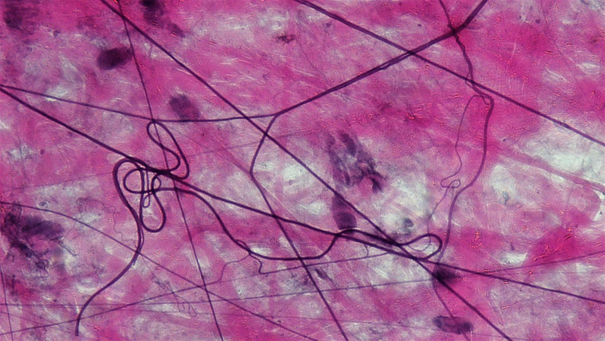
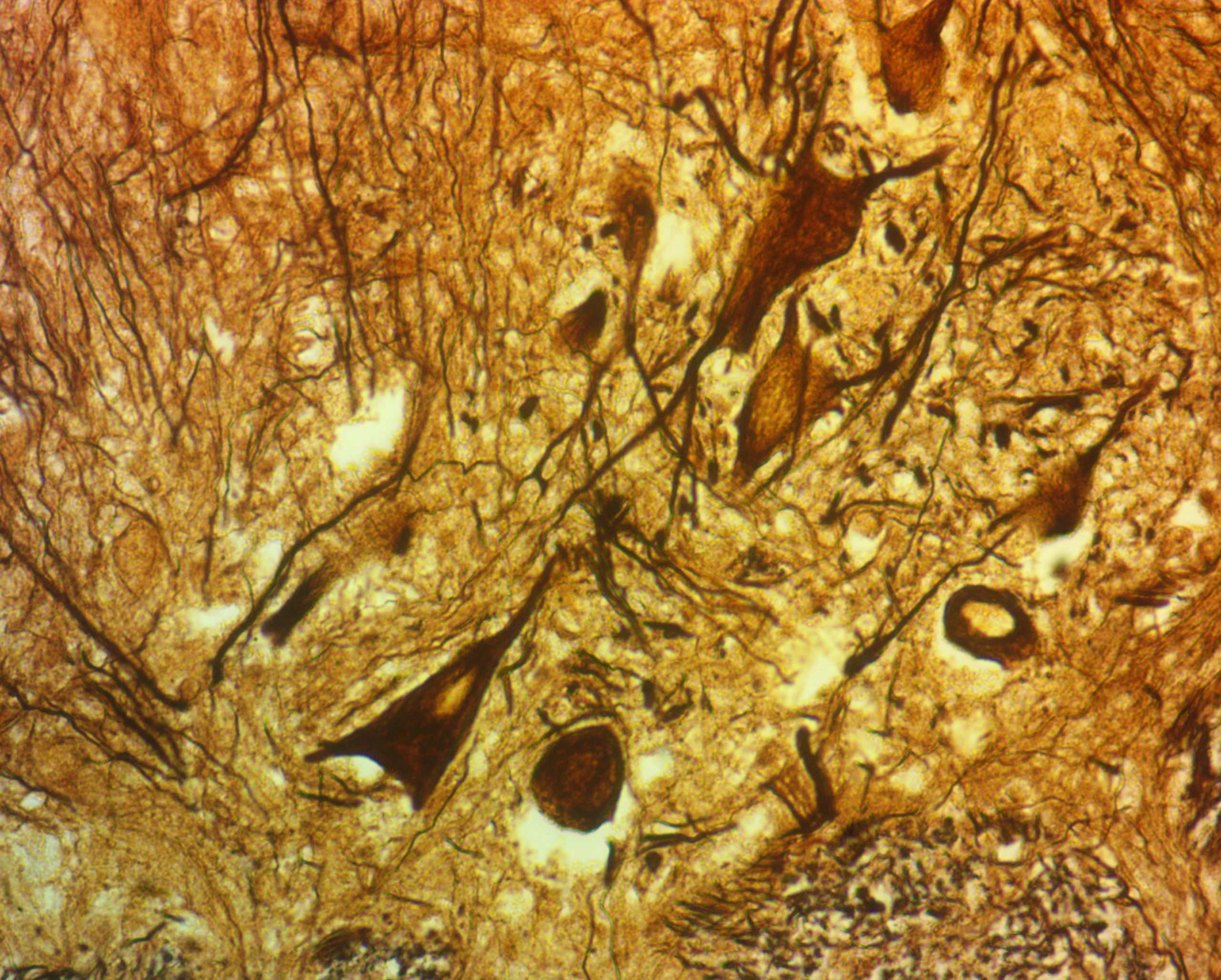
- Move slide horizontally and vertically to locate neurons. They are large and blue (or dark brown) in color with no definite shape. Dendrites and axons are extensions of the cytoplasm and appear like hair. Locate dendrites that are numerous and thin. Axons are thicker and fewer in number.
- Change magnification to high power (10 x 10). Notice neuroglial cells in cytoplasm are dense in number.
- Draw what you observed at low and high magnification, showing neuronal cell bodies, glial cells, axons, dendrites.
Exercise 5. Histologically Impaired Tissues
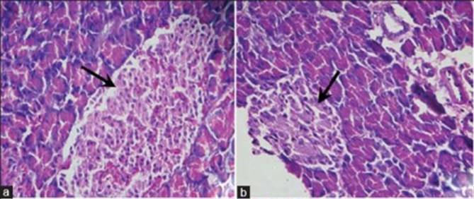
At the beginning of this chapter on Tissues, we referred to diabetes. The organ that is affected is the pancreas. Notice changes in the structure of the pancreas by comparing it with normal tissue. (Figure 4.13)
This is what you should do:
- Ask your instructor for a pancreas slide.
- Place it on the stage of the microscope and bring the tissue into focus using low power (4 x 10) magnification.
- Move slide horizontally and vertically to locate the Islets of Langerhans, acini and ducts (Figure 4.1 and 4.13).
- Change magnification to high power (10 x 10). Notice the epithelial cells in the islets and acini
- Draw what you observed for this normal pancreas tissue at low and high magnification, showing islet and acinar epithelial cells.
- How is the normal pancreas different than the diabetic pancreas in Figure 4.13? Describe the differences in the Isles of Langerhans between the normal and diabetic specimens.
Post-Laboratory Questions
- What type of cells make up epithelial tissue? Sketch the cell to show the three different shapes that are found in epithelial tissues.
- Epithelial tissue is characterized by (a) shape and (b) layers. Name one tissue to demonstrate shape as an example.
- Compare lung tissue and kidney tissue. How are they different? How are they similar?
- Sketch a pseudostratified epithelium. What is its basic characteristic?
- Give one example each of a solid, semi-solid and fluid connective tissue. State the characteristics that define each tissue.
- How are the three types of muscle tissues different from each other?
- How would you identify a neuron if you saw one?
