
3 Chapter 3 The Cellular Level of Organization
By Krishnan Prabhakaran
The Immortal Woman : Henrietta Lacks and HeLa Cells
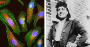
Figure 3.1 HeLa Cells: A Lasting Contribution to Biomedical Research (Credit: National Institutes of Health, Office of Science Policy. https://osp.od.nih.gov/scientific-sharing/hela-cells-landing/)
Motivation. In 1951, Henrietta Lacks, a 31-year-old African-American woman, went to Baltimore’s Johns Hopkins Hospital to be treated for cervical cancer. Some of her cancer cells began being used in research due to their unique ability to continuously grow and divide in the laboratory. These so-called “immortal” cells were later named “HeLa” after the first two letters of Henrietta Lacks first and last name. Since Ms. Lacks’ untimely death in 1952, HeLa cells have been a vital tool in biomedical research, leading to an increased understanding of the fundamentals of human health and disease. Some of the research involving HeLa cells also served as the underpinning of several Nobel Prize winning discoveries.
While Henrietta Lacks’ story has been known in the research community for some time, it raised further awareness after the publication of the best-selling book The Immortal Life of Henrietta Lacks (Crown, 2010). To honor Ms. Lacks’ and her family’s continued support of biomedical research, NIH analyzed and evaluated the scientific literature involving HeLa cells and found over 110,000 publications that cited the use of HeLa cells between 1953 to 2018. This analysis further highlights the persistent impact of HeLa cells in science and medicine, proving that they have been a consistent, essential tool that has allowed researchers to expand the knowledge base in fields such as cancer biology, infectious disease, and many others.
Learning Objectives
Upon completion of the work in this chapter, students should be able to:
- Define cell & organelle
- Identify major regions of the cell (cytoplasm, nucleus, plasma membrane) on a model and/or diagram
- Summarize major functions of each major region of the cell.
- Identify typical organelles found in an animal cell and summarize major functions of each
- Summarize the major events for each part of the cell cycle (interphase and mitosis)
Background
All living matter is composed of cells. The human body contains trillions of cells. The metabolism of living organisms, all their biochemical activities, takes place within cells and as a result of cellular activity. All cells arise from other cells by cell division. All cells are surrounded by a cell membrane which encloses the cytoplasm (protoplasm) and various other internal structures. The cell membrane restricts passage of materials in and out of the cell and helps to protect the cells structural and functional integrity. Internally, floating in the cytoplasm, are various organelles (small organs), each with a specific function similar to some of the organs found in our bodies. A nucleus is found in almost all of our cells and is often the largest cellular structure present. The nucleus contains the genetic material, the chromosomes, which are made of DNA and control all metabolism. Most cells also contain mitochondria which contain most of the enzymes for extracting energy from organic foods, a chemical process called respiration.
The Cell Membrane
Despite differences in structure and function, all living cells in multicellular organisms have a surrounding cell membrane. As the outer layer of your skin separates your body from its environment, the cell membrane (also known as the plasma membrane) separates the inner contents of a cell (intracellular) from its exterior environment (extracellular). The cell membrane provides a protective barrier around the cell and regulates which materials can pass in or out.
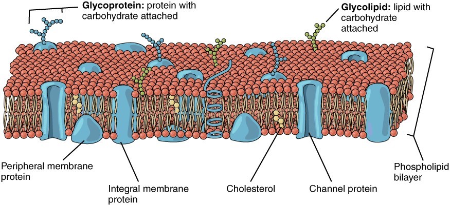
The cell membrane is an extremely pliable and variable cell structure composed primarily of back-to-back phospholipids (the phospholipid bilayer). Cholesterol is also present, which contributes to the fluidity of the membrane, and there are various proteins embedded within the membrane that have a variety of functions. The two major structural classes of proteins are integral proteins and peripheral proteins. Integral proteins are embedded into the cell membrane and allow cells to move materials between the intracellular and extracellular environments and communicate with other cells. A channel protein is an example of an integral protein that selectively allows particular materials, such as certain ions, to pass into or out of the cell. Peripheral proteins are typically found on the inner or outer surface of the lipid bilayer but can also be attached to the internal or external surface of an integral protein. These proteins typically perform a specific function for the cell and this includes proteins that act as digestive enzymes to break down nutrients in the small intestine so that they are small enough to be absorbed by the cells.
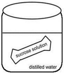
Diffusion and Osmosis
Diffusion is the process by which molecules spread from areas of high concentration to areas of low concentration. This movement, down the concentration gradient, continues until molecules are evenly distributed. Osmosis is a special type of diffusion: the diffusion of water through a semipermeable membrane. The concentration of water is inversely related to the concentration of solute: more solute corresponds to less water and less solute corresponds to more water. This is important because osmotic vocabulary describes the solute and not the water. Hypertonic solutions contain a high concentration of solute and little water, relative to hypotonic solutions that have a low concentration of solute and therefore a higher concentration of water. The term “isotonic” is used when two areas have an equal concentration of solute: no net osmosis is occurring (Credit: biologycorner.com).
The Cytoplasm
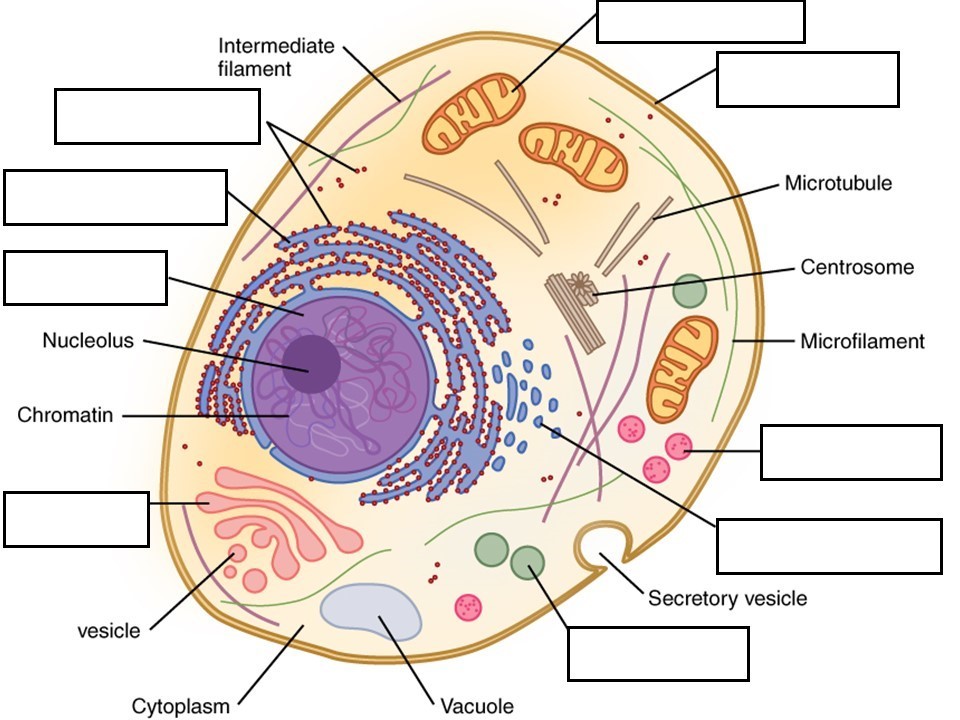
All living cells in multicellular organisms contain an internal compartment, called the cytoplasm which includes the cytosol, organelles, and the cytoskeleton. The cytosol is the fluid component of the cytoplasm and is a jelly-like substance within the cell that includes the components necessary for cellular function. Cells also contain various membrane-enclosed cellular organelles which perform a specific function and are described further below. The cytoskeleton is a group of fibrous proteins, including microfilaments, intermediate filaments, and microtubules that helps cells maintain their structural integrity. Cytoskeletal components are also critical for cell motility, cell reproduction, and transportation of substances within the cell.
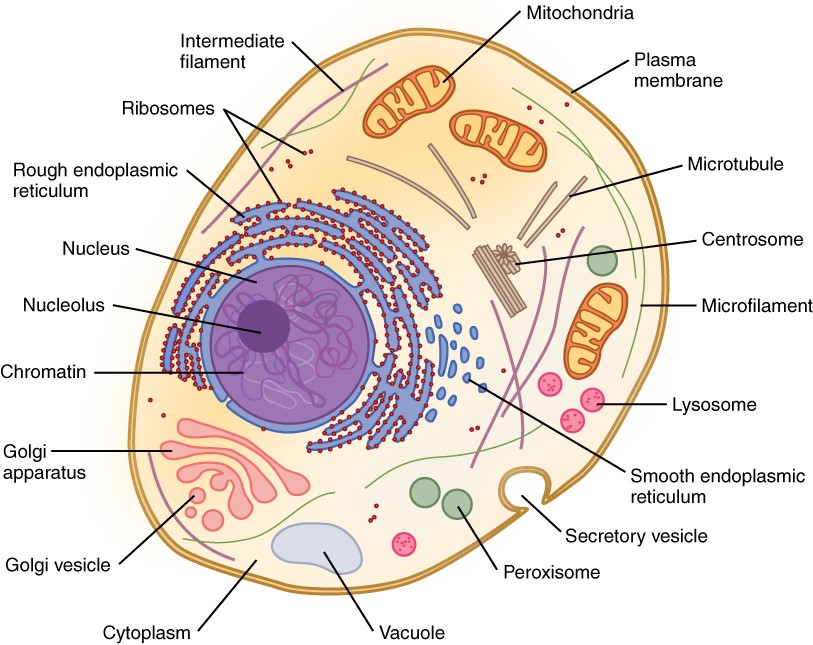
Organelles
Just as the various organs work together in harmony to perform all of a human’s necessary functions, cellular organelles work together to keep the cell performing all of its important functions. Figure 3.2 shows several examples of cellular organelles whose function is summarized in Table 3.1.
| Table 3.1 | |
| Organelle | Function |
| Nucleus | Contains the cell’s DNA and directs cellular functions. |
| Mitochondrion | Converts energy storage molecules into the major energy molecule, ATP, to power cellular function |
| Ribosome | Protein synthesis |
| Rough endoplasmic reticulum | Includes ribosomes for the synthesis and modification of proteins |
| Smooth endoplasmic reticulum | Lipid synthesis |
| Golgi apparatus | Sorts, modifies, and ships products from the endoplasmic reticulum |
| Lysosome | Contains digestive enzymes to break down materials |
| Peroxisome | Contains enzymes key for lipid metabolism and chemical detoxification |
The Cell Cycle
The cell cycle consists of two general phases: interphase, followed by mitosis and cytokinesis (Figure 3.4). Interphase is the period of the cell cycle during which the cell is not dividing. The majority of cells are in interphase most of the time. Mitosis is the division of genetic material, during which the cell nucleus breaks down and two new, fully functional, nuclei are formed. Cytokinesis divides the cytoplasm into two distinctive cells.
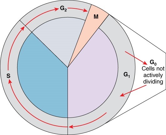
Interphase
A cell grows and carries out all normal metabolic functions and processes in a period called G1 (Figure 3.4). G1 phase (gap 1 phase) is the first gap, or growth phase in the cell cycle and is the phase that varies the most in terms of duration. Cells might spend a couple of hours, or many days in this phase. For cells that will divide again, G1 is followed by replication of the DNA, during S phase. S phase (synthesis phase) is the period during which a cell replicates its DNA. After S phase, the cell proceeds through the G2 phase. The G2 phase is a second gap phase, during which the cell continues to grow and makes the necessary preparations for mitosis. S phase typically lasts between 8-10 hours and the G2 phase approximately 5 hours. Cells that have temporarily stopped dividing and are resting (a common state) and cells that have permanently ceased dividing (like nerve cells) are said to be in G0, a resting phase of the cell cycle.
Mitosis and Cytokinesis
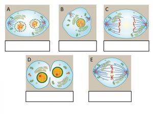
The mitotic phase of the cell cycle typically takes between 1 and 2 hours. During this phase, a cell undergoes two major processes. First, it completes mitosis, during which the contents of the nucleus are equitably pulled apart and distributed between its two halves. Cytokinesis then occurs, dividing the cytoplasm and cell body into two new cells. Mitosis is divided into four major stages that take place after interphase (Figure 3.5) in the following order: prophase, metaphase, anaphase, and telophase. The process is then followed by cytokinesis
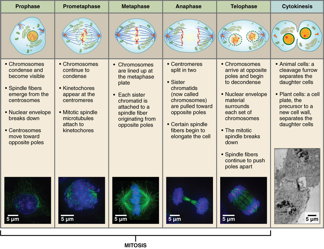
Prophase
Prophase is the first phase of mitosis, during which the loosely packed chromatin coils and condenses into visible chromosomes. During prophase, each chromosome becomes visible with its identical partner attached, forming the familiar X-shape of sister chromatids. The nucleolus disappears early during this phase, and the nuclear envelope also disintegrates. Centrosomes migrate to two different sides of the cell and microtubules begin to extend from each like long fingers from two hands extending toward each other.
Metaphase
Metaphase is the second stage of mitosis. During this stage, the sister chromatids, with their attached microtubules, line up along a linear plane in the middle of the cell. A metaphase plate forms between the centrosomes that are now located at either end of the cell. Microtubules are now poised to pull apart the sister chromatids and bring one from each pair to each side of the cell.
Anaphase
Anaphase is the third stage of mitosis. Anaphase takes place over a few minutes, when the pairs of sister chromatids are separated from one another, forming individual chromosomes once again. Each end of the cell receives one partner from each pair of sister chromatids, ensuring that the two new daughter cells will contain identical genetic material.
Telophase
Telophase is the final stage of mitosis. Telophase is characterized by the formation of two new daughter nuclei at either end of the dividing cell. These newly formed nuclei surround the genetic material, which uncoils such that the chromosomes return to loosely packed chromatin. Nucleoli also reappear within the new nuclei, and the mitotic spindle breaks apart, each new cell receiving its own complement of DNA, organelles, membranes, and centrioles. At this point, the cell is already beginning to split in half as cytokinesis begins.
Cytokinesis
The cleavage furrow is a contractile band made up of microfilaments that forms around the midline of the cell during cytokinesis. This contractile band squeezes the two cells apart until they finally separate. Two new cells are now formed.
Microscopy
A microscope is an instrument that magnifies an object so that it may be seen by the observer. Because cells are usually too small to see with the naked eye, a microscope is an essential tool in the field of biology. In addition to magnification, microscopes also provide resolution, which is the ability to distinguish two nearby objects as separate. A combination of magnification and resolution is necessary to clearly view specimens under the microscope. The light microscope bends a beam of light at the specimen using a series of lenses to provide a clear image of the specimen to the observer.
In this lab, parts of the microscope will be reviewed. Students will learn proper use and care of the microscope.
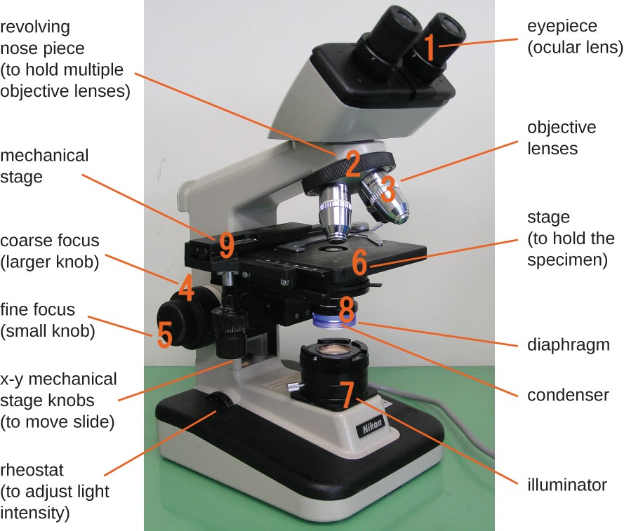
Magnification
Your microscope has 4 objective lenses: Scanning (4x), Low (10x), High (40x), and Oil Immersion (100x). In addition to the objective lenses, the ocular lens (eyepiece) has a magnification. The total magnification is determined by multiplying the magnification of the ocular and objective lenses. The total magnification is the product of the ocular magnification times the objective magnification:
For example, if a 40⨯ objective lens is selected and the ocular lens is 10⨯, the total magnification would be
(40×)(10×)=400×
General Procedures
- Make sure all backpacks, purses, etc. are off the benchtop.
- Carry microscope by the base and arm with both hands.
- Store with cord wrapped around microscope and the scanning objective clicked into place.
Focusing Specimens
- Plug your microscope in to power supply and switch on illuminator.
- Always start with the stage as low as possible and using scanning objective (4x). Odds are, you will be able to see something on this setting (sometimes it’s only a color). Use the coarse knob to focus: the image may be small at this magnification, but you won’t be able to find it on the higher powers without this first step. Move the mechanical stage until your focused image is also centered.
- Once you’ve focused using the scanning objective, switch to the low power objective (10x). Use the coarse knob to refocus and move the mechanical stage to re-center your image. Again, if you haven’t focused on this level, you will not be able to move to the next level.
- Now switch to the high power objective (40x). At this point, ONLY use the fine adjustment knob to focus specimens.
- If the specimen is too light or too dark, try adjusting the diaphragm.
Cleanup
- Store microscope with the scanning objective in place and the stage in its lowest position.
- Wrap cords around microscope. Replace slides to original slide tray
- Replace slides to original slide tray.
Pre-Laboratory Questions
1. Which of the following is/are found in cell membranes? Select all that apply.
A. Carbohydrates
B. Phospholipids
C. Proteins
D. Cholesterol
E. All of the above are associated with the membrane
2. Which type of lipid is most important in biological membranes?
A. Waxes
B. Fats
C. Oils
D. Triglycerides
E. Phospholipids
3. In facilitated diffusion, molecules move through pores from areas of ____________ concentration to areas of ____________ concentration.
A. high, high
B. low, high
C. high, low
4. What structure moves the chromosomes to the correct location for cell division?
A. Microtubule
B. Nucleus
C. Centriole
D. Chromatids
Exercises
- Exercise 1. Identification of cell components on a cell model (optional)
- Exercise 2. Observing cells with a compound microscope
- Exercise 3. Diffusion through a gel
- Exercise 4. Osmosis across a membrane
- Exercise 5. Cell anatomy (optional)
- Exercise 6. Cell cycle (optional)
Exercise 1 Identification of cell components on a model (optional)
Required Materials
- Classroom model of an animal cell
- Post-it notes
Procedure
Study the cell model. Identify the following organelles and structures, and place a post-it note on the model to identify the structure. Additionally, label the cell figure below with the correct structure names.
-
Peroxisome Smooth ER Ribosome Nucleus Mitochondrion Plasma membrane Lysosome Golgi apparatus Rough ER
Exercise 2 Observing cells with a compound microscope
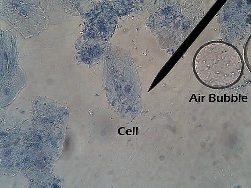
Prepare a wet mount of a human cheek cell and observe under the microscope.
Required Materials
- Compound microscope
- Methylene blue
- Microscope slide
- Coverslip
- Toothpick
Procedure
- Put a drop of methylene blue on the slide.
- Gently scrape the inside of your cheek with the flatside of a toothpick. Scrape lightly!
- Stir the end of the toothpick in the methylene blue stain on the slide.
- Discard the toothpick in a trash container labeled “biohazard waste”.
- Place a coverslip onto the slide.
- Place the prepared wet mount microscope sample onto the stage of a microscope.
- Use the SCANNING (4x) objective to focus. You probably will not see the cells at this power.
- Switch to low power (10x). Cells should be visible, but they will be small and look like nearly clear purplish blobs. If you are looking at something very dark purple, it is probably not a cell.
- Once you think you have located a cell, switch to high power (40x) and refocus. Remember, do NOT use the coarse adjustment knob at this point!
- Sketch the cell at low and high power. Label the nucleus, cytoplasm, and cell membrane of a single cell
Exercise 3 Diffusion through a gel
One factor that can affect the rate of diffusion is the size of the molecule. Larger molecules tend to move more slowly than smaller molecules. In this experiment, students will compare the diffusion rates of two dyes traveling through agar.
Required Materials
- Pre-punched agar plates
- Potassium permanganate
- Janus green
- Ruler (cm)
Procedure
- Using the dropper, drop a single drop of potassium permanganate into one of the wells on the plate.
- Repeat with Janus green.
- Allow the plates to sit undisturbed for 30 minutes.
- Which dye do you think will have the faster diffusion rate? ________________
- After 30 minutes, measure the radius of the dye front from the middle of the well and record your results.
- Calculate the diffusion rate (mm/hr) by dividing the dye front radius by 0.5.
| Potassium permanganate | Janus Green | |
| Molecular Weight | 158g/mole | 511g/mole |
| Radius (mm) | ||
| Diffusion Rate |
Questions:
- Did your outcome match your expectation? Provide an explanation for your results.
- What are other factors that can affect the rate of diffusion?
Exercise 4 Osmosis across a membrane
Observe the movement of water across a semipermeable membrane.
Required Materials
- Dialysis bags (4 per group)
- Dental floss
- 15% sucrose solution
- 30% sucrose solution
- Triple beam balance
- Beakers
- Graduated cylinder
- Stir rods
- Water bath
Procedure
- Place 4 strips of dialysis tubing in a water bath until the tubing softens enough to open it up.
- Tie a tight knot in one end of each dialysis tubing / bag using the dental floss.
- Pour approximately 10 mL of each solution into separate bags (see table below).
- Fill out the table indicating the Tonicity of Bag (isotonic, hypotonic, hypertonic) compared to the beaker.
- Indicate whether you expect water to move into or out of the bag under Predicted Outcome.
Tube 1 Tube 2 Tube 3 Tube 4 Inside bag water 15% sucrose 30% sucrose water Inside beaker water water water 15% sucrose Tonicity of Bag Predicted Outcome - Remove most of the air from the bag (but leave a little bit of space) and tie the bag on the other end.
- Blot the bags to remove any substances that may have spilled; check the bags for leaks.
- Record the weight of each dialysis bag/tube in the data table as Weight at 0 min.
Data Tube 1 Tube 2 Tube 3 Tube 4 Weight at 0 min Weight at 10 min Weight at 20 min Weight at 30 min - Place a bag/tube in each beaker (be sure to keep track of which bag is in which beaker!). Fill the beakers with enough of the appropriate solution to cover your bags (refer to the above table).
- Predict what you think will happen during the experiment.
- Record baggie weight every 10 minutes in data table.
- After 30 minutes, remove the bags from solution and record the final weight.
Exercise 5 Cell Anatomy (optional)
Required Materials
- None
Procedure
This activity will be completed individually or in small groups. Refer to the background information as needed to help answer the questions below.
Complete the table below to summarize the major function of each given organelle in one sentence or phrase.
| Organelle | Function (one sentence or phrase) |
| Peroxisome | |
| Nucleus | |
| Lysosome | |
| Smooth ER | |
| Mitochondrion | |
| Golgi apparatus | |
| Ribosome | |
| Rough ER |
Exercise 6 Cell Cycle (optional)
Required Materials
- Classroom model of Mitosis
Procedure
This activity will be completed individually or in small groups. Refer to the background information as needed to help answer the questions below.
A. Match the cell cycle phase to the major cellular events by completing table below with the provided cell cycle phases. Each cell cycle phase will only be used once.
S phase, G1 phase, Anaphase, Prophase, Metaphase, Telophase, Cytokinesis, G0 phase
| Cell Cycle Phase | Major Cellular Events |
| Sister chromatids are pulled to opposite poles | |
| Sister chromatids line up at middle of cell | |
| Chromosomes de-condense and nuclear envelopes reform | |
| Cleavage furrow separates daughter cells | |
| Significant cell growth to prepare for mitosis | |
| Replication of DNA | |
| Condensation of chromosomes | |
| Cells are not dividing |
B. Label each of the following drawings of cells in different stages of mitosis and cytokinesis
Post-laboratory Questions
1. Nemo is a clownfish and clownfish live in the ocean. The salinity of the Nemo’s ocean environment averages around 3.5%. What would happen to Nemo if he became lost and ended up in a fresh water lake?
A. Nemo would die because his cells would lose water and he would dehydrate quickly.
B. Nemo would swell and could pop if he doesn’t get out of there.
C. Nemo would be fine, there wouldn’t be any water movement
2. The chemical transmitter dopamine is released from a vesicle within a neuron when the vesicle fuses with the cell membrane and dumps the chemical into the extracellular space. Which type of transport mechanism is most representative of this process?
A. active transport
B. diffusion
C. exocytosis
D. endocytosis
3. The solution in beaker A is 20% of glucose and the solution in beaker B is 40% of glucose, which beaker is hypertonic?
A. beaker A
B. beaker B
4. If you connected the beakers with a tube that was permeable to water but impermeable to glucose, which way would water move?
A. From beaker A to beaker B
B. From beaker B to beaker A
C. Water would not move
